Passa al contenuto
Merck
Tutte le immagini(2)

Documenti fondamentali

MABC604

Sigma-Aldrich

Anti-MLKL Antibody, clone 3H1

clone 3H1, from rat

Sinonimo/i:

Mixed lineage kinase domain-like protein, MLKL

Autenticatiper visualizzare i prezzi riservati alla tua organizzazione & contrattuali


About This Item

Codice UNSPSC:
12352203
eCl@ss:
32160702
NACRES:
NA.41

Origine biologica

rat

Livello qualitativo

Forma dell’anticorpo

purified antibody

Tipo di anticorpo

primary antibodies

Clone

3H1, monoclonal

Reattività contro le specie

mouse

tecniche

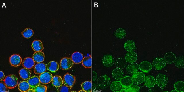
immunocytochemistry: suitable
immunoprecipitation (IP): suitable
western blot: suitable

Isotipo

IgG

N° accesso NCBI

N° accesso UniProt

Condizioni di spedizione

wet ice

modifica post-traduzionali bersaglio

unmodified

Informazioni sul gene

mouse ... Mlkl(74568)

Descrizione generale

MLKL, also known as Mixed lineage kinase domain-like protein, and encoded by the gene MLKL, is an interesting protein that is required for the execution of programmed necrosis or necroptosis, for example in response to TNF-alpha induced necrosis. MLKL is a component of the "necrosome," the multiprotein complex that triggers tumor necrosis factor (TNF)-induced cell death by necroptosis. The programmed necrosis induced by TNF-α also requires the activities of the receptor-interacting serine-threonine kinases RIP1 and RIP3 and they too interact directly with the mixed lineage kinase domain-like protein MLKL. RIP3 is an essential upstream kinase in necroptosis. The pseudokinase MLKL functions as a substrate of RIP3 to mediate downstream signaling and when complexed to MLKL the RIP3-MLKL complex is stabilized by AMP-PNP to adopt an inactive conformation.

Immunogeno

Epitope: Brace region.
KLH-conjugated linear peptide corresponding to a sequence from the two-helix linker (Brace) region N-terminal to the pseudokinase domain of mouse MLKL (Hildebrand, J.M., et al. (2014). Proc. Natl. Acad. Sci. U.S.A. 111(42):15072-15077; Murphy, J.M., et al. (2013). Immunity. 39(3):443-453).

Applicazioni

Immunocytochemistry Analysis: A representative lot detected MLKL in mouse dermal fibroblasts (MDFs) from wild-type, but not Mlkl-/- mice (Courtesy of Prof. James M. Murphy, Walter and Eliza Hall Institute, Australia).

Immunoprecipitation Analysis: A representative lot immunoprecipitated MLKL from soluble extract of Mlkl-/- mouse dermal fibroblasts (MDFs) harboring doxycycline-inducible wild-type mouse MLKL expression construct only after, but not before doxycycline treatment (Murphy, J.M., et al. (2013). Immunity. 39(3):443-453).

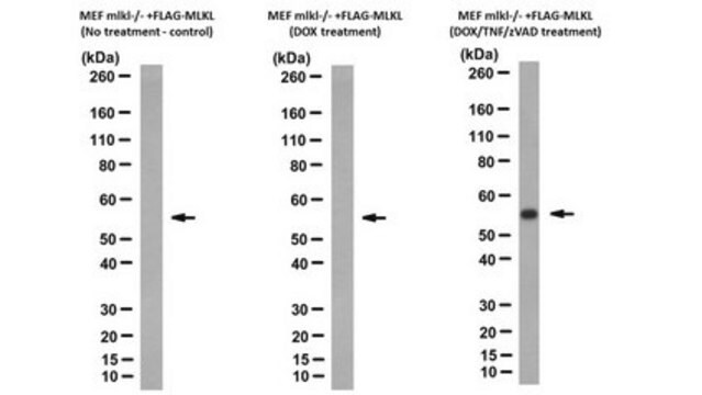
Western Blotting Analysis: A representative lot detected Q-VD-OPh (TSQ; Cat. No. 551476) treatment-induced membrane translocation of murine and equine MLKL N-terminal fragment (a.a. 1-180 and 1-189, respectively) exogenously expressed in mouse dermal fibroblasts (MDFs) from Mlkl-/- mice (Tanzer, M.C., et al. (2016). Cell Death Differ.. In press).

Western Blotting Analysis: A representative lot detected MLKL in human HT-29 and U937 cells (Tanzer, M.C., et al. (2015). Biochem. J. 471(2):255-265).

Western Blotting Analysis: Representative lots detected MLKL membrane translocation in mouse dermal fibroblasts (MDFs) upon Q-VD-OPh (TSQ; Cat. No. 551476) treatment (Tanzer, M.C., et al. (2015). Biochem. J. 471(2):255-265; Hildebrand, J.M., et al. (2014). Proc. Natl. Acad. Sci. U.S.A. 111(42):15072-15077).

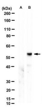
Western Blotting Analysis: Representative lots detected MLKL expression in L292 mouse fibroblasts, as well as in mouse dermal fibroblasts (MDFs), mouse embryonic fibroblasts (MEFs) and bone marrow derived macrophages (BMDMs) from wild-type, but not Mlkl-/- mice (Cook, W.D., et al. (2014). Cell Death Differ. 21(10):1600-1612; Murphy, J.M., et al. (2013). Immunity. 39(3):443-453).

Western Blotting Analysis: A representative lot detected recombinant full-length mouse MLKL as well as a.a. 1-180 and a.a. 124-464, but not a.a. 1-125 or a.a. 179-464, MLKL fragments (Hildebrand, J.M., et al. (2014). Proc. Natl. Acad. Sci. U.S.A. 111(42):15072-15077).

Western Blotting Analysis: A representative lot detected MLKL expression in all tissues tested except brain from wild-type mice, while no target band was seen in any tissues from Mlkl-/- mice (Murphy, J.M., et al. (2013). Immunity. 39(3):443-453).
This Anti-MLKL Antibody, clone 3H1 is validated for use in Immunocytochemistry, Immunoprecipitation, and Western Blotting for the detection of MLKL.

Qualità

Evaluated by Western Blotting in mouse heart tissue lysate.

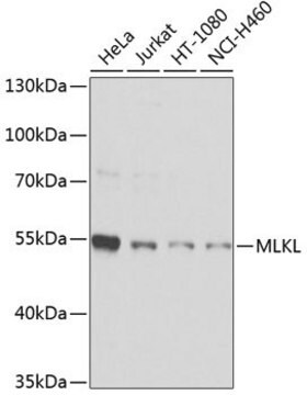
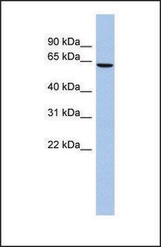
Western Blotting Analysis: 0.5 µg/mL of this antibody detected MLKL in 10 µg of mouse heart tissue lysate.

Descrizione del bersaglio

~52 kDa observed. 54.48/30.28 (human isoform 1/2) and 54.32/53.38 kDa (mouse isoform 1/2) calculated. Uncharacterized band(s) may be observed in some cell lysates.

Stato fisico

Format: Purified
Purified rat IgG in buffer containing 0.1 M Tris-Glycine (pH 7.4), 150 mM NaCl with 0.05% sodium azide.

Altre note

Concentration: Please refer to the Certificate of Analysis for the lot-specific concentration.

Non trovi il prodotto giusto?  

Prova il nostro Motore di ricerca dei prodotti.

Raccomandato

N° Catalogo
Descrizione
Determinazione del prezzo

Codice della classe di stoccaggio

12 - Non Combustible Liquids

Classe di pericolosità dell'acqua (WGK)

WGK 1

Punto d’infiammabilità (°F)

Not applicable

Punto d’infiammabilità (°C)

Not applicable


Certificati d'analisi (COA)

Cerca il Certificati d'analisi (COA) digitando il numero di lotto/batch corrispondente. I numeri di lotto o di batch sono stampati sull'etichetta dei prodotti dopo la parola ‘Lotto’ o ‘Batch’.

Possiedi già questo prodotto?

I documenti relativi ai prodotti acquistati recentemente sono disponibili nell’Archivio dei documenti.

Visita l’Archivio dei documenti

The pseudokinase MLKL mediates necroptosis via a molecular switch mechanism.
Murphy, James M, et al.
Immunity, 39, 443-453 (2013)
RIPK1- and RIPK3-induced cell death mode is determined by target availability.
Cook, WD; Moujalled, DM; Ralph, TJ; Lock, P; Young, SN; Murphy, JM; Vaux, DL
Cell Death and Differentiation null
Snahel Patel et al.
Cell death and differentiation, 27(1), 161-175 (2019-05-19)
The kinase RIP1 acts in multiple signaling pathways to regulate inflammatory responses and it can trigger both apoptosis and necroptosis. Its kinase activity has been implicated in a range of inflammatory, neurodegenerative, and oncogenic diseases. Here, we explore the effect
Joseph Sarhan et al.
Cell death and differentiation, 26(2), 332-347 (2018-05-23)
Interferons (IFNs) are critical determinants in immune-competence and autoimmunity, and are endogenously regulated by a low-level constitutive feedback loop. However, little is known about the functions and origins of constitutive IFN. Recently, lipopolysaccharide (LPS)-induced IFN was implicated as a driver
Hayley I Muendlein et al.
Cell reports, 30(3), 699-713 (2020-01-23)
Receptor-interacting protein kinase 1 (RIPK1) and 3 (RIPK3) are well known for their capacity to drive necroptosis via mixed-lineage kinase-like domain (MLKL). Recently, RIPK1/3 kinase activity has been shown to drive inflammation via activation of MAPK signaling. However, the regulatory

Il team dei nostri ricercatori vanta grande esperienza in tutte le aree della ricerca quali Life Science, scienza dei materiali, sintesi chimica, cromatografia, discipline analitiche, ecc..

Contatta l'Assistenza Tecnica.