Skip to Content
Merck
All Photos(2)

Documents

MABE78

Sigma-Aldrich

Anti-MiTF Antibody, clone 3D1.2

clone 3D1.2, from mouse

Synonym(s):

Microphthalmia-associated transcription factor, Class E basic helix-loop-helix protein 32, bHLHe32

Sign Into View Organizational & Contract Pricing


About This Item

UNSPSC Code:
12352203
eCl@ss:
32160702
NACRES:
NA.41

biological source

mouse

Quality Level

antibody form

purified immunoglobulin

antibody product type

primary antibodies

clone

3D1.2, monoclonal

species reactivity

human, mouse

technique(s)

immunohistochemistry: suitable
western blot: suitable

isotype

IgMκ

NCBI accession no.

UniProt accession no.

shipped in

wet ice

target post-translational modification

unmodified

Gene Information

human ... MITF(4286)

General description

Microphthalmia-associated transcription factor (MiTF) is a basic helix loop helix leucine zipper (b HLH ZIP) transcription factor implicated in pigmentation, and development of mast cells and bone. Mutations in MiTF cause auditory pigmentary syndromes in humans, such as Waardenburg syndrome type II and type IIa, and Tietz syndrome. There are two known isoforms of MiTF, differing by 66 amino acids at the N-terminus. Shorter forms are expressed in melanocytes and run as two bands at 52 kDa and 56 kDa, whereas the longer Mi form runs as a cluster of bands at 60-70 kDa in osteoclasts and in B16 melonoma cells (but not other melanoma cell lines), as well as mast cells and heart. MiTF plays a critical role in the differentiation of various cell types as neural crest-derived melanocytes, mast cells, osteoclasts and optic cup-derived retinal pigment epithelium.

Immunogen

GST-tagged recombinant protein corresponding to human MiTF.

Application

Anti-MiTF Antibody, clone 3D1.2 is an antibody against MiTF for use in Western Blotting & IHC.
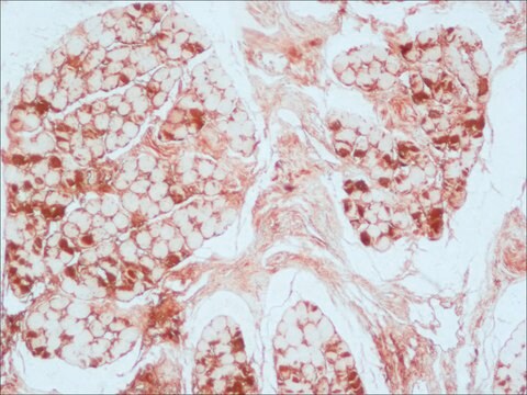
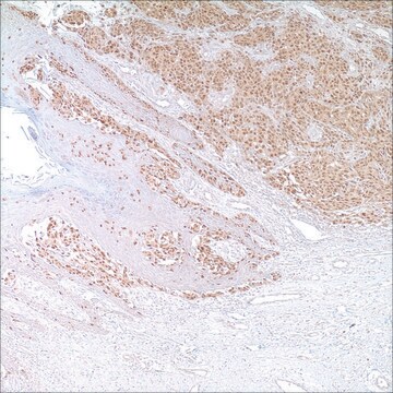
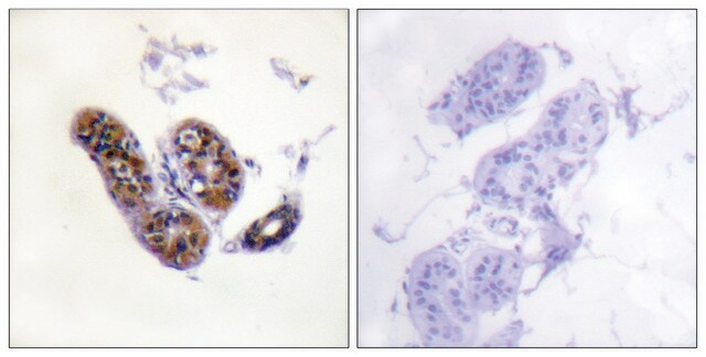
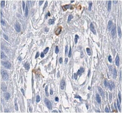
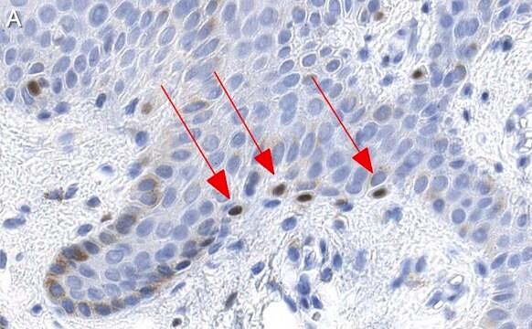
Immunohistochemistry Analysis: A 1:100 dilution of this antibody detected MiTF in human melanoma tissue.
Research Category
Signaling
Research Sub Category
Transcription Factors

Quality

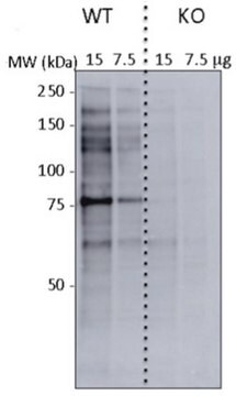
Evaluated by Western Blot in mouse brain tissue lysate.

Western Blot Analysis: A 1:500 dilution of this antibody detected MiTF in 10 µg of mouse brain tissue lysate.

Target description

~52 kDa observed. Multiple isoforms between 46-56 kDa are known to exist.

Linkage

Replaces: MAB3747

Physical form

Format: Purified
Purified mouse monoclonal IgMκ in buffer containing 0.01M PBS, 0.1% sodium azide, pH 7.1.

Storage and Stability

Stable for 1 year at 2-8°C from date of receipt.

Analysis Note

Control
Mouse brain tissue lysate

Disclaimer

Unless otherwise stated in our catalog or other company documentation accompanying the product(s), our products are intended for research use only and are not to be used for any other purpose, which includes but is not limited to, unauthorized commercial uses, in vitro diagnostic uses, ex vivo or in vivo therapeutic uses or any type of consumption or application to humans or animals.

Not finding the right product?  

Try our Product Selector Tool.

Storage Class Code

12 - Non Combustible Liquids

WGK

WGK 1

Flash Point(F)

Not applicable

Flash Point(C)

Not applicable


Certificates of Analysis (COA)

Search for Certificates of Analysis (COA) by entering the products Lot/Batch Number. Lot and Batch Numbers can be found on a product’s label following the words ‘Lot’ or ‘Batch’.

Already Own This Product?

Find documentation for the products that you have recently purchased in the Document Library.

Visit the Document Library

Our team of scientists has experience in all areas of research including Life Science, Material Science, Chemical Synthesis, Chromatography, Analytical and many others.

Contact Technical Service