Accéder au contenu
Merck
Toutes les photos(1)

Principaux documents

MABN819

Sigma-Aldrich

Anti-Tau Antibody, oligomeric Antibody, clone TOMA-1

clone TOMA-1, from mouse

Synonyme(s) :

Microtubule-associated protein tau oligomer, Tau oligomer, PHF-tau oligomer, Paired helical filament-tau oligomer, Neurofibrillary tangle protein oligomer

Se connecterpour consulter vos tarifs contractuels et ceux de votre entreprise/organisme


About This Item

Code UNSPSC :
12352203
eCl@ss :
32160702
Nomenclature NACRES :
NA.41

Source biologique

mouse

Niveau de qualité

Forme d'anticorps

purified immunoglobulin

Type de produit anticorps

primary antibodies

Clone

TOMA-1, monoclonal

Espèces réactives

rat, human, mouse

Réactivité de l'espèce (prédite par homologie)

mammals (based on high homology)

Technique(s)

ELISA: suitable
dot blot: suitable
immunofluorescence: suitable
immunohistochemistry: suitable
neutralization: suitable
western blot: suitable

Isotype

IgG2aκ

Numéro d'accès NCBI

Numéro d'accès UniProt

Conditions d'expédition

ambient

Modification post-traductionnelle de la cible

unmodified

Informations sur le gène

human ... MAPT(4137)
mouse ... Mapt(17762)
rat ... Mapt(29477)

Description générale

Microtubule-associated protein tau (UniProt P10636; also known as Neurofibrillary tangle protein, Paired helical filament-tau, PHF-tau) is encoded by the MAPT (also known as TAU, MAPT1, MTBTL) gene (Gene ID 4137) in human. In Alzheimer′s disease (AD) pathology, accumulation of the microtubule-associated protein tau takes place primarily in the neurons. Tau accumulates in both the somatodendritic and axonal domains of neurons. Tau also accumulates in the soma as neurofibrillary tangles (NFTs). Cell death and synaptic lesions occur independently of NFT formation, and research indicates that NFT formation alone is insufficient for neurodegeneration, suggesting that soluble tau aggregates may be the more toxic and pathologically significant tau species. Tau oligomers are neurotoxic when applied extracellularly to cultured neuronal cells, and tau oligomers (but not fibrils) induce neurodegeneration and synaptic and mitochondrial dysfunction in vivo. Moreove, researchers are able to use tau oligomers as a reliable biomarker to differentiate AD brains from age-matched non-AD brains.

Spécificité

Clone TOMA-1 (a.k.a. clone H12C10) is among more than 13 hybridomas derived from mice immunized with in vitro generated recombinant Tau-441 (Tau-F, Tau-4, 2N4R isoform) oligomers. These TOMAs are reported to specifically detect oligomeric tau without any significant reactivity toward monomeric tau, tau fibrils, Aβ oligomers, Aβ fibrils, or α-synuclein oligomers (Castillo-Carranza, D.L., et al. (2014). J. Neurosci. 34(12):4260-4272).

Immunogène

Recombinant human Tau-441 (Tau-F, Tau-4, 2N4R isoform) oligomers (Lasagna-Reeves, C. A., et al. (2012). FASEB J. 26(5):1946-1959).

Application

Anti-Tau, oligomeric, clone TOMA-1, Cat. No. MABN819, is amouse monoclonal antibody that targets tau oligomers and has been tested in Dot Blot, ELISA, Immunofluorescence, Immunohistochemistry, Neutralization, and Western Blotting applications.
Research Category
Neuroscience
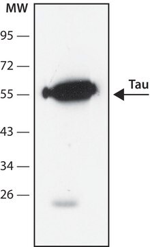
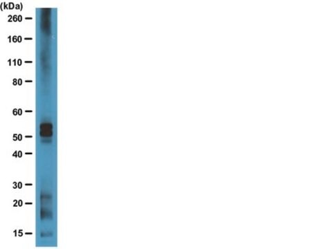
Western Blotting Analysis: 20 µg/mL from a representative lot detected upregulated oligomeric tau in soluble PBS brain extract from Alzheimer′s diseased (AD) human brain (Courtesy of Sengupta, U., et al., Kayed lab, University of Texas, Galveston).

Please refer to the following publications regarding the use of TOMA clones in Dot Blot, ELISA, Immunofluorescence, Immunohistochemistry, Neutralization, and Western Blotting applications:

1. Vuono, R., et al. (2015). Brain. 138(Pt 7):1907-1918.
2. Castillo-Carranza, D.L., et al. (2015). J. Neurosci. 35(12):4857-4868.
3. Castillo-Carranza, D.L., et al. (2014). J. Neurosci. 34(12):4260-4272.

Qualité

Evaluated by Western Blotting in Alzheimer′s diseased human brain lysate.

Western Blotting Analysis: 4 µg/mL of this antibody detected Tau oligmers in 25 µg of Alzheimer′s diseased human brain lysate.

Description de la cible

Variable depending on the size(s) of the oligomer(s). Uncharacterized bands may be observed in some lysate(s).

Forme physique

Format: Purified
Protein A purified.
Purified mouse IgG2aκ in buffer containing PBS without preservatives.

Stockage et stabilité

Stable for 1 year at -20°C from date of receipt.

Autres remarques

Concentration: Please refer to lot specific datasheet.

Clause de non-responsabilité

Unless otherwise stated in our catalog or other company documentation accompanying the product(s), our products are intended for research use only and are not to be used for any other purpose, which includes but is not limited to, unauthorized commercial uses, in vitro diagnostic uses, ex vivo or in vivo therapeutic uses or any type of consumption or application to humans or animals.

Vous ne trouvez pas le bon produit ?  

Essayez notre Outil de sélection de produits.

Code de la classe de stockage

12 - Non Combustible Liquids

Classe de danger pour l'eau (WGK)

WGK 2

Point d'éclair (°F)

Not applicable

Point d'éclair (°C)

Not applicable


Certificats d'analyse (COA)

Recherchez un Certificats d'analyse (COA) en saisissant le numéro de lot du produit. Les numéros de lot figurent sur l'étiquette du produit après les mots "Lot" ou "Batch".

Déjà en possession de ce produit ?

Retrouvez la documentation relative aux produits que vous avez récemment achetés dans la Bibliothèque de documents.

Consulter la Bibliothèque de documents

Fan Xia et al.
Acta neuropathologica communications, 9(1), 51-51 (2021-03-26)
The retina, as the only visually accessible tissue in the central nervous system, has attracted significant attention for evaluating it as a biomarker for neurodegenerative diseases. Yet, most of studies focus on characterizing the loss of retinal ganglion cells (RGCs)
Marius Chiasseu et al.
Neurobiology of disease, 145, 105059-105059 (2020-08-29)
There is a strong unmet need for translational progress towards Alzheimer's disease (AD) modifying therapy. Unfortunately, preclinical modeling of the disease has been disappointing, relying primarily on transgenic mouse overexpression of rare dominant mutations. Clinical manifestation of AD symptoms is
Shiyao Wang et al.
Biotechnology progress, 36(1), e2920-e2920 (2019-10-04)
Microtubule-associated protein tau is an intrinsically disordered, highly soluble protein found primarily in neurons. Under normal conditions, tau regulates the stability of axonal microtubules and intracellular vesicle transport. However, in patients of neurodegeneration such as Alzheimer's disease (AD), tau forms

Notre équipe de scientifiques dispose d'une expérience dans tous les secteurs de la recherche, notamment en sciences de la vie, science des matériaux, synthèse chimique, chromatographie, analyse et dans de nombreux autres domaines..

Contacter notre Service technique