Saltar al contenido
Merck

P8874

Sigma-Aldrich

Monoclonal Anti-Phosphocan antibody produced in mouse

~2 mg/mL, clone 122.2, purified immunoglobulin, buffered aqueous solution

Sinónimos:

Anti-PTPRB, Anti-Receptor-type Protein-tyrosine phosphatase β

Iniciar sesiónpara Ver la Fijación de precios por contrato y de la organización


About This Item

Número MDL:
Código UNSPSC:
12352203
NACRES:
NA.41

origen biológico

mouse

Nivel de calidad

conjugado

unconjugated

forma del anticuerpo

purified immunoglobulin

tipo de anticuerpo

primary antibodies

clon

122.2, monoclonal

formulario

buffered aqueous solution

mol peso

antigen ~180 kDa (higher band may be present)

reactividad de especies

rat

envase

antibody small pack of 25 μL

concentración

~2 mg/mL

técnicas

immunocytochemistry: suitable
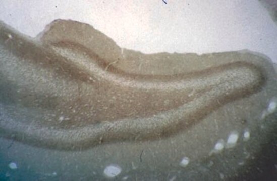
immunohistochemistry: suitable
western blot: 0.2-0.4 μg/mL using total extract of rat brain

isotipo

IgM

Nº de acceso UniProt

Condiciones de envío

dry ice

temp. de almacenamiento

−20°C

modificación del objetivo postraduccional

unmodified

Información sobre el gen

Descripción general

Monoclonal Anti-Phosphacan (mouse IgM isotype) is derived from the hybridoma 122.2 produced by the fusion of mouse myeloma cells (P3X cells) and splenocytes from BALB/c mice immunized with rat brain proteoglycans. Chondroitin sulfate proteoglycans are neural cell adhesion molecules (NCAM) ligands present in the brain extracellular matrix (ECM). Phosphacan protein is expressed mainly in astrocytes and is a ligand for NCAM. Phosphacan is the soluble extracellular domain of the receptor-type transmembrane protein tyrosine phosphatase (RPTPb).

Inmunógeno

rat brain proteoglycans.

Aplicación

Monoclonal Anti-Phosphacan antibody produced in mouse has been used in:
  • immunoblotting
  • immunohistochemistry
  • immunocytochemistry.

Acciones bioquímicas o fisiológicas

Phosphacan levels elevates during late embryogenesis. It reaches a plateau two weeks postnatal before reaching stable. Receptor-type transmembrane protein tyrosine phosphatase (RPTPb) functions to promote primary tecal neurons neurite growth, neural migration and also induces cell adhesion. Both phosphacan and RPTPb can bind to NCAM and tenascin-C and −R. Phosphacan can oppose RPTPb by competing for its binding sites. Both in hippocampal and spinal cord neurons, phosphacan can affect neuronal adhesion and neurite outgrowth.

Forma física

Solution in 0.01 M phosphate buffered saline, pH 7.4, containing 15 mM sodium azide.

Cláusula de descargo de responsabilidad

Unless otherwise stated in our catalog or other company documentation accompanying the product(s), our products are intended for research use only and are not to be used for any other purpose, which includes but is not limited to, unauthorized commercial uses, in vitro diagnostic uses, ex vivo or in vivo therapeutic uses or any type of consumption or application to humans or animals.

¿No encuentra el producto adecuado?  

Pruebe nuestro Herramienta de selección de productos.

Código de clase de almacenamiento

10 - Combustible liquids

Clase de riesgo para el agua (WGK)

WGK 3

Punto de inflamabilidad (°F)

Not applicable

Punto de inflamabilidad (°C)

Not applicable

Equipo de protección personal

Eyeshields, Gloves, multi-purpose combination respirator cartridge (US)


Certificados de análisis (COA)

Busque Certificados de análisis (COA) introduciendo el número de lote del producto. Los números de lote se encuentran en la etiqueta del producto después de las palabras «Lot» o «Batch»

¿Ya tiene este producto?

Encuentre la documentación para los productos que ha comprado recientemente en la Biblioteca de documentos.

Visite la Librería de documentos

Receptor protein tyrosine phosphatases in nervous system development
Johnson KG and Van Vactor D
Physiological Reviews, 83(1), 1-24 (2003)
The tissue plasminogen activator (tPA/Plasmin) extracellular proteolytic system regulates seizure-induced hippocampal mossy fiber outgrowth through a proteoglycan substrate
Wu YP, et al.
The Journal of cell biology, 148(6), 1295-1304 (2000)
Y P Wu et al.
The Journal of cell biology, 148(6), 1295-1304 (2000-03-22)
Short seizure episodes are associated with remodeling of neuronal connections. One region where such reorganization occurs is the hippocampus, and in particular, the mossy fiber pathway. Using genetic and pharmacological approaches, we show here a critical role in vivo for
RGMa mediates reactive astrogliosis and glial scar formation through TGFbeta1/Smad2/3 signaling after stroke
Zhang R, et al.
Cell Death and Differentiation, 25(8), 1503-1503 (2018)
Rongrong Zhang et al.
Cell death and differentiation, 25(8), 1503-1516 (2018-02-06)
In response to stroke, astrocytes become reactive astrogliosis and are a major component of a glial scar. This results in the formation of both a physical and chemical (production of chondroitin sulfate proteoglycans) barrier, which prevent neurite regeneration that, in

Nuestro equipo de científicos tiene experiencia en todas las áreas de investigación: Ciencias de la vida, Ciencia de los materiales, Síntesis química, Cromatografía, Analítica y muchas otras.

Póngase en contacto con el Servicio técnico