Saltar al contenido
Merck
Todas las fotos(1)

Documentos clave

358M-1

Sigma-Aldrich

MUM1 (MRQ-8) Mouse Monoclonal Antibody

Iniciar sesiónpara Ver la Fijación de precios por contrato y de la organización


About This Item

Código UNSPSC:
12352203
NACRES:
NA.41

origen biológico

mouse

Nivel de calidad

100
500

conjugado

unconjugated

forma del anticuerpo

culture supernatant

tipo de anticuerpo

primary antibodies

clon

MRQ-8, monoclonal

descripción

For In Vitro Diagnostic Use in Select Regions (See Chart)

Formulario

buffered aqueous solution

reactividad de especies

human

envase

vial of 0.1 mL concentrate (358M-14)
vial of 0.5 mL concentrate (358M-15)
bottle of 1.0 mL predilute (358M-17)
vial of 1.0 mL concentrate (358M-16)
bottle of 7.0 mL predilute (358M-18)

fabricante / nombre comercial

Cell Marque

técnicas

immunohistochemistry (formalin-fixed, paraffin-embedded sections): 1:100-1:500

isotipo

IgG1κ

control

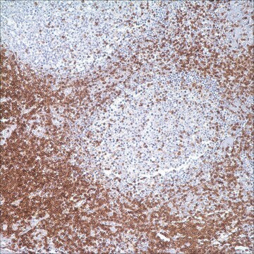
B-cell chronic lymphocytic leukemia (B-CLL), plasma cell myeloma, tonsil

Condiciones de envío

wet ice

temp. de almacenamiento

2-8°C

visualización

cytoplasmic, nuclear

Información sobre el gen

human ... PWWP3A(84939)

Descripción general

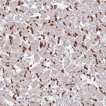
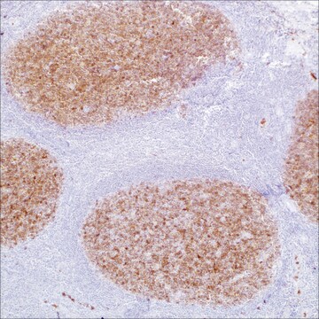
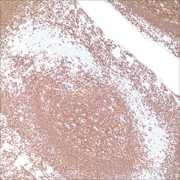
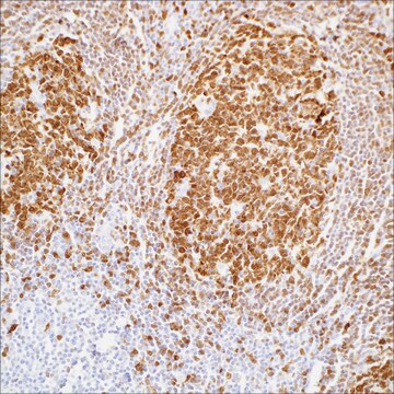
MUM1 (multiple myeloma oncogene-1)/IRF4 (interferon regulatory factor 4) is a 50 kDa protein encoded by MUM1 gene, and a member of the interferon regulatory factor family of transcription factors. MUM1/IRF4 is expressed in the nuclei of plasma cells and a small percentage of germinal center (GC) B-cells located in the “light zone”. This antibody stains MUM1 protein, which is expressed in a subset of B-cells in the light zone of the germinal center, plasma cells, activated T-cells, and a wide spectrum of related hematolymphoid neoplasms derived from these cells. Therefore, this antibody is useful for the subclassiffication of lymphoid malignancies and an excellent marker for Reed-Sternberg cells of classic Hodgkin disease in combination with anti-CD30.

Calidad


IVD

IVD

IVD

RUO

Ligadura / enlace

MUM1 Positive Control Slides, Product No. 358S, are available for immunohistochemistry (formalin-fixed, paraffin-embedded sections).

Forma física

Solution in Tris Buffer, pH 7.3-7.7, with 1% BSA and <0.1% Sodium Azide

Nota de preparación

Download the IFU specific to your product lot and formatNote: This requires a keycode which can be found on your packaging or product label.

Otras notas

For Technical Service please contact: 800-665-7284 or email: service@cellmarque.com

Información legal

Cell Marque is a trademark of Merck KGaA, Darmstadt, Germany

¿No encuentra el producto adecuado?  

Pruebe nuestro Herramienta de selección de productos.


Elija entre una de las versiones más recientes:

Certificados de análisis (COA)

Lot/Batch Number

¿No ve la versión correcta?

Si necesita una versión concreta, puede buscar un certificado específico por el número de lote.

¿Ya tiene este producto?

Encuentre la documentación para los productos que ha comprado recientemente en la Biblioteca de documentos.

Visite la Librería de documentos

G Gaidano et al.
Leukemia, 14(4), 563-566 (2000-04-14)
In recent times, the field of B cell lymphoma histogenesis has progressed rapidly due to the increasing availability of histogenetic markers. Genotypic markers of B cell histogenesis are represented by mutations of IgV and BCL-6 genes, which are somatically acquired
Y Natkunam et al.
Modern pathology : an official journal of the United States and Canadian Academy of Pathology, Inc, 14(7), 686-694 (2001-07-17)
The gene encoding MUM1 was characterized as a possible translocation partner in chromosomal abnormalities involving a significant number of multiple myelomas. The overexpression of the MUM1 protein as a result of translocation t(6;14) (p25;q32) identified MUM1 as a putative regulatory
B Falini et al.
Blood, 95(6), 2084-2092 (2000-03-09)
A new monoclonal antibody (MUM1p) was used to study the cell/tissue expression of human MUM1/IRF4 protein, the product of the homologous gene involved in the myeloma-associated t(6;14) (p25;q32). MUM1 was expressed in the nuclei and cytoplasm of plasma cells and
Antonino Carbone et al.
British journal of haematology, 117(2), 366-372 (2002-04-26)
Biological and clinical studies have shown that Hodgkin's disease (HD) can be divided into two major categories, termed nodular lymphocyte predominance HD (NLP HD) and classic HD (CHD). Within CHD four subtypes have been distinguished: nodular sclerosis, mixed cellularity, lymphocyte
Mathewos Tessema et al.
PloS one, 7(4), e34850-e34850 (2012-04-13)
Aberrant cytosine methylation affects regulation of hundreds of genes during cancer development. In this study, a novel aberrantly hypermethylated CpG island in cancer was discovered within the TOX2 promoter. TOX2 was unmethylated in normal cells but 28% lung (n =

Nuestro equipo de científicos tiene experiencia en todas las áreas de investigación: Ciencias de la vida, Ciencia de los materiales, Síntesis química, Cromatografía, Analítica y muchas otras.

Póngase en contacto con el Servicio técnico