
MABT851M
Anti-Podoplanin Antibody, clone PMab-1
clone PMab-1, from rat
Sinónimos:
Aggrus, Glycoprotein 38, Gp38, OTS-8, PA2.26 antigen, T1-alpha, T1A, Transmembrane glycoprotein E11
About This Item
Productos recomendados


origen biológico
rat
Nivel de calidad
forma del anticuerpo
purified antibody
tipo de anticuerpo
primary antibodies
clon
PMab-1, monoclonal
reactividad de especies
mouse
envase
antibody small pack of 25 μg
técnicas
flow cytometry: suitable
immunocytochemistry: suitable
immunohistochemistry: suitable (paraffin)
western blot: suitable
isotipo
IgG2aκ
Nº de acceso NCBI
Nº de acceso UniProt
modificación del objetivo postraduccional
unmodified
Información sobre el gen
mouse ... Pdpn(14726)
Descripción general
Especificidad
Inmunógeno
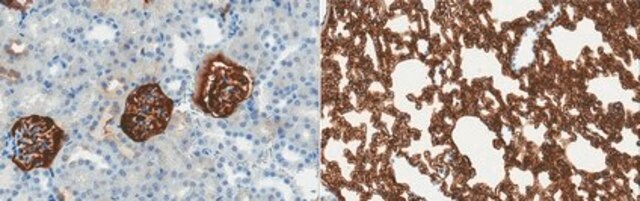
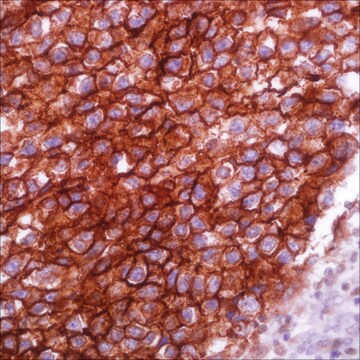
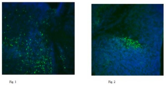
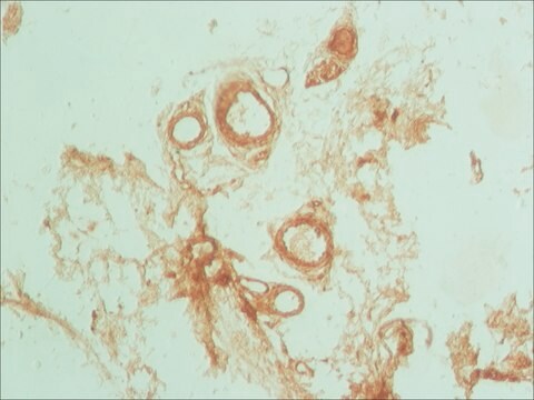
Aplicación
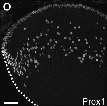
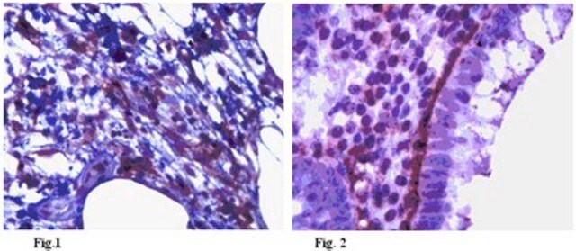
Immunohistochemistry Analysis: A representative lot detected Podoplanin in Immunohistochemistry applications (Kaji, C., et. al. (2012) Acta Histochem Cytochem. 45(4):227-37).
Flow Cytometry Analysis: A representative lot detected Podoplanin in Flow Cytometry applications (Oki, H., et. al. (2015). Monoclon Antib Immunodiagn Immunother. 34(6):396-403).
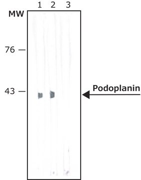
Western Blotting Analysis: A representative lot detected Podoplanin in Western Blotting applications (Oki, H., et. al. (2015). Monoclon Antib Immunodiagn Immunother. 34(6):396-403).
Immunocytochemistry Analysis: A representative lot detected Podoplanin in Immunocytochemistry applications (Kaji, C., et. al. (2012) Acta Histochem Cytochem. 45(4):227-37).
Cell Structure
Calidad
Western Blotting Analysis: 1 µg/mL of this antibody detected Podoplanin in 10 µg of mouse colon tissue lysate.
Descripción de destino
Forma física
Almacenamiento y estabilidad
Otras notas
Cláusula de descargo de responsabilidad
¿No encuentra el producto adecuado?
Pruebe nuestro Herramienta de selección de productos.
Código de clase de almacenamiento
12 - Non Combustible Liquids
Clase de riesgo para el agua (WGK)
WGK 1
Punto de inflamabilidad (°F)
Not applicable
Punto de inflamabilidad (°C)
Not applicable
Certificados de análisis (COA)
Busque Certificados de análisis (COA) introduciendo el número de lote del producto. Los números de lote se encuentran en la etiqueta del producto después de las palabras «Lot» o «Batch»
¿Ya tiene este producto?
Encuentre la documentación para los productos que ha comprado recientemente en la Biblioteca de documentos.
Nuestro equipo de científicos tiene experiencia en todas las áreas de investigación: Ciencias de la vida, Ciencia de los materiales, Síntesis química, Cromatografía, Analítica y muchas otras.
Póngase en contacto con el Servicio técnico