Descripción general
Cytokeratin-8/18 (UniProt: P05787/P05783; also known as Keratin, type II cytoskeletal 8/ Keratin, type I cytoskeletal 18, Keratin-8/Keratin-18, K8/K18) is encoded by the KRT8/KRT18 (also known as CYK8/CYK18) gene (Gene ID: 3856/3875) in human. Cytokeratins 8 and 18 (K8/18) are simple epithelial cell-specific intermediate filament proteins. Cytokeratin-8 is a type II, neutral to basic, protein of the intermediate filament family that together with Cytokeratin-19 (KRT19) helps to link the contractile apparatus to dystrophin at the costameres of striated muscle. Cytokeratin-8 can undergo phosphorylation on three major serine residues: Serine 23, 431, and 73. Serine 23 is shown to be highly conserved in all type II keratins. Phosphorylation at Serine 73 is reported to increase during cellular stress, including hear and drug exposure. However, under normal conditions serine 73 remains largely dephosphorylated. Cytokeratin-8 can also undergo O-glycosylation in a cell cycle-dependent manner and glycosylation increases its solubility and reduces stability by inducing proteasomal degradation. Cytokeratin-18 is a type I, acidic, protein that is expressed in colon, placenta, and liver. Higher expression levels have been reported in lymph nodes of breast carcinoma. Cytokeratin-18 is involved in the uptake of thrombin-antithrombin complexes by hepatic cells. Upon phosphorylation, it plays a role in filament reorganization. It is proteolytically cleaved by caspases during epithelial cell apoptosis and this cleavage is shown to occur at Asp-238. Phosphorylation of cytokeratin-18 at serine 33 is shown to increase during mitosis in cultured cells and in regenerating liver. Serine 33 phosphorylation is considered to be essential for its association with 14-3-3 proteins and has a role in keratin organization and distribution. Cytokeratin-18 can undergo O-glycosylation, which results in its increased solubility and reduction in its stability. Mutations in KRT8 and KRT18 genes have been been linked to liver cirrhosis that is characterized by severe panlobular liver-cell swelling with Mallory body formation, prominent pericellular fibrosis, and marked deposits of copper.
Especificidad
Clone L2A1 is a mouse monoclonal antibody that detects human cytokeratin 8/18.
Inmunógeno
KLH-conjugated Linear Peptide corresponding to a sequence from Human Cytokeratin 8/18.
Aplicación
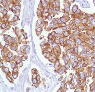
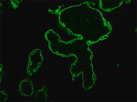
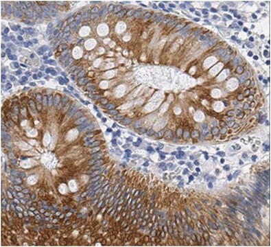
Anti-Cytokeratin 8/18, clone L2A1, Cat. No. MABT844, is a mouse monoclonal antibody that detects cytokeratin 8/18 and has been tested for use in Immunocytochemistry and Immunoprecipitation.
Immunoprecipitation Analysis: A representative lot detected Cytokeratin 8/18 in Immunoprecipitation applications (Haltiwanger, R.S., et. al. (1997). J Biol Chem. 272(13):8752-8; Chou, C.F., et. al. (1994). Biochem J. 298 ( Pt 2):457-63; Chou, C.F., et. al. (1993). Biochem J. 268(6):4465-72; Ku, N.O., et. al. (2000). J Cell Biol. 149(3):547-52; Lahdeniemi, I.A.K., et. al. (2017). Cell Death Differ. 24(6):984-996).
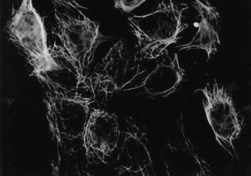
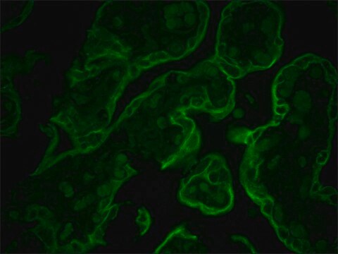
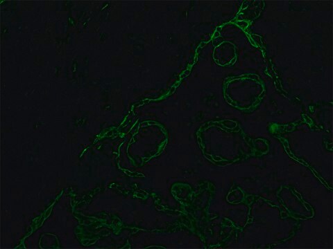
Immunocytochemistry Analysis: A representative lot detected Cytokeratin 8/18 in Immunocytochemistry applications (Chou, C.F., et. al. (1993). Biochem J. 268(6):4465-72).
Research Category
Cell Structure
Calidad
Evaluated by Immunocytochemistry in HT-29 cells.
Immunocytochemistry Analysis: A 1:500 dilution of this antibody detected Cytokeratin 8/18 in HT-29 cells.
Descripción de destino
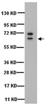
~53.70 kDa and 48.06 kDa, respectively for cytokeratin 8 and 18 calculated. Uncharacterized bands may be observed in some lysate(s).
Forma física
Format: Purified
Protein G purified
Purified mouse monoclonal antibody IgG in buffer containing 0.1 M Tris-Glycine (pH 7.4), 150 mM NaCl with 0.05% sodium azide.
Almacenamiento y estabilidad
Stable for 1 year at 2-8°C from date of receipt.
Otras notas
Concentration: Please refer to lot specific datasheet.
Cláusula de descargo de responsabilidad
Unless otherwise stated in our catalog or other company documentation accompanying the product(s), our products are intended for research use only and are not to be used for any other purpose, which includes but is not limited to, unauthorized commercial uses, in vitro diagnostic uses, ex vivo or in vivo therapeutic uses or any type of consumption or application to humans or animals.
