Saltar al contenido
Merck
Todas las fotos(2)

Key Documents

MABN2406

Sigma-Aldrich

Anti-Tau Antibody, clone TNT-2

clone TNT-2, from mouse

Sinónimos:

Microtubule-associated protein tau, Neurofibrillary tangle protein, Paired helical filament-tau, PHF-tau

Iniciar sesiónpara Ver la Fijación de precios por contrato y de la organización


About This Item

Código UNSPSC:
12352203
eCl@ss:
32160702

origen biológico

mouse

Nivel de calidad

forma del anticuerpo

purified immunoglobulin

tipo de anticuerpo

primary antibodies

clon

TNT-2, monoclonal

reactividad de especies

rat, human, mouse

técnicas

ELISA: suitable
immunofluorescence: suitable
immunohistochemistry: suitable
western blot: suitable

isotipo

IgG1κ

Nº de acceso NCBI

Nº de acceso UniProt

Condiciones de envío

ambient

modificación del objetivo postraduccional

unmodified

Información sobre el gen

human ... MAPT(4137)
mouse ... Mapt(17762)
rat ... Mapt(29477)

Descripción general

Microtubule-associated protein tau (UniProt: P10636; also known as Neurofibrillary tangle protein, Paired helical filament-tau, PHF-tau) is encoded by the MAPT (MAPTL, MTBT1, TAU) gene (Gene ID: 4137) in human. Tau proteins are mostly found in the axons of neurons, in the cytosol and in association with plasma membrane components. Normally, Tau promotes microtubule assembly and stability, and might be involved in the establishment and maintenance of neuronal polarity. The C-terminus binds axonal microtubules while the N-terminus binds neural plasma membrane components, suggesting that tau functions as a linker protein between both. Axonal polarity is predetermined by TAU/MAPT localization (in the neuronal cell) in the domain of the cell body defined by the centrosome. The localization of Tau protein in the Alzheimer s disease (AD) brain is markedly abnormal and may contribute to neuronal dysfunction. In AD, normal soluble cytoskeletal elements, such as Tau and neurofilaments, are transformed into insoluble paired helical filaments (PHFs). This is linked to the post-translational change in Tau, primarily its hyperphosphorylation by a number of protein kinases. In its non-phosphorylated state, Tau promotes rapid and extensive microtubule (MT) polymerization. A significant consequence of Tau hyperphosphorylation in AD is a reduction in its ability to bind microtubules and promote microtubule assembly. Hyperphosphorylated Tau may contribute to a destabilized microtubule network, impaired axonal transport, and ultimately result in NFT formation and neuronal death.

Especificidad

Clone TNT-2 specirfically detects tau in human, mouse, and rat brain. It targets a sequence of 17 amino acids from the N-terminal region that is common to all isoforms (1-6) of tau.

Inmunógeno

KLH-conjugated linear peptide corresponding to 17 amino acids from the N-terminal region of human Tau.

Aplicación

Anti-Tau Antibody, clone TNT-2, Cat. No. MABN2406, is a highly specific mouse monoclonal antibody that targets Tau and has been tested in ELISA, Immunofluorescence, Immunohistochemistry, and Western Blotting.
Research Category
Neuroscience
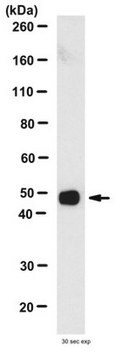
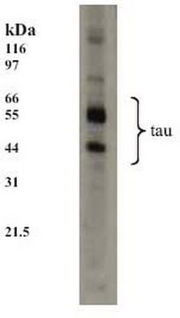
Western Blotting Analysis: 0.02 µg/mL from a representative lot detected Tau in 10 µg of mouse entorhinal cortex.

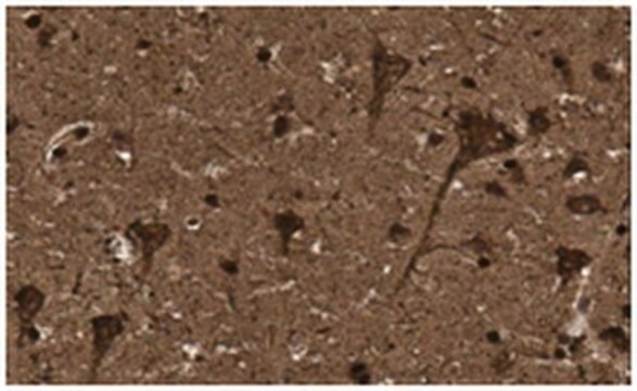
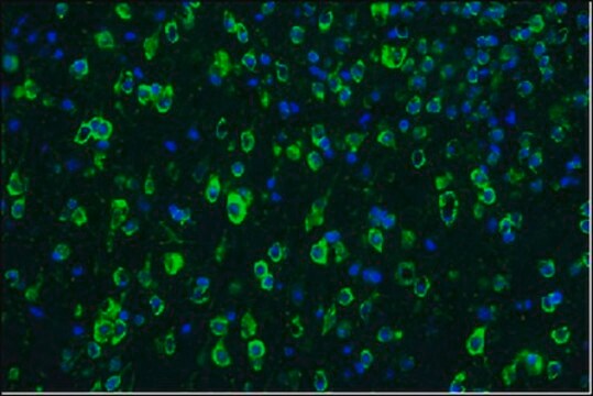
Immunohistochemistry Analysis: A representative lot detected Tau in Immunohistochemistry applications (Combs, B., et. al. (2016). Neurobiol Dis. 94:18-31).

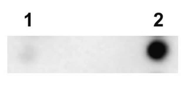
ELISA Analysis: A representative lot detected Tau in ELISA applications (Combs, B., et. al. (2016). Neurobiol Dis. 94:18-31).

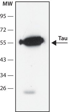
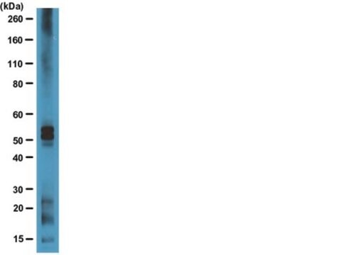
Western Blotting Analysis: A representative lot detected Tau in Western Blotting applications (Combs, B., et. al. (2016). Neurobiol Dis. 94:18-31).

Immunofluorescence Analysis: A representative lot detected Tau in Immunofluorescence applications (Combs, B., et. al. (2016). Neurobiol Dis. 94:18-31).

Calidad

Evaluated by Western Blotting in rat entorhinal cortex.

Western Blotting Analysis: 0.02 µg/mL of this antibody detected Tau in 10 µg of rat entorhinal cortex lysate.

Descripción de destino

~55 kda Observed. Uncharacterized bands may be observed in some lysate(s).

Forma física

Format: Purified
Protein G purified
Purified mouse monoclonal antibody IgG1 in buffer containing 0.1 M Tris-Glycine (pH 7.4), 150 mM NaCl with 0.05% sodium azide.

Almacenamiento y estabilidad

Stable for 1 year at 2-8°C from date of receipt.

Otras notas

Concentration: Please refer to lot specific datasheet.

Cláusula de descargo de responsabilidad

Unless otherwise stated in our catalog or other company documentation accompanying the product(s), our products are intended for research use only and are not to be used for any other purpose, which includes but is not limited to, unauthorized commercial uses, in vitro diagnostic uses, ex vivo or in vivo therapeutic uses or any type of consumption or application to humans or animals.

¿No encuentra el producto adecuado?  

Pruebe nuestro Herramienta de selección de productos.

Código de clase de almacenamiento

12 - Non Combustible Liquids

Clase de riesgo para el agua (WGK)

WGK 1

Punto de inflamabilidad (°F)

Not applicable

Punto de inflamabilidad (°C)

Not applicable


Certificados de análisis (COA)

Busque Certificados de análisis (COA) introduciendo el número de lote del producto. Los números de lote se encuentran en la etiqueta del producto después de las palabras «Lot» o «Batch»

¿Ya tiene este producto?

Encuentre la documentación para los productos que ha comprado recientemente en la Biblioteca de documentos.

Visite la Librería de documentos

Nuestro equipo de científicos tiene experiencia en todas las áreas de investigación: Ciencias de la vida, Ciencia de los materiales, Síntesis química, Cromatografía, Analítica y muchas otras.

Póngase en contacto con el Servicio técnico