Saltar al contenido
Merck
Todas las fotos(1)

Documentos clave

MABF958

Sigma-Aldrich

Anti-CD47 Antibody, clone PF3.1

clone PF3.1, from mouse

Sinónimos:

Leukocyte surface antigen CD47, Antigenic surface determinant protein OA3, CD47, IAP, Integrin-associated protein, Protein MER6

Iniciar sesiónpara Ver la Fijación de precios por contrato y de la organización


About This Item

Código UNSPSC:
12352203
eCl@ss:
32160702
NACRES:
NA.41

origen biológico

mouse

Nivel de calidad

forma del anticuerpo

purified immunoglobulin

tipo de anticuerpo

primary antibodies

clon

PF3.1, monoclonal

reactividad de especies

human

técnicas

flow cytometry: suitable

isotipo

IgG1κ

Nº de acceso NCBI

Nº de acceso UniProt

Condiciones de envío

ambient

modificación del objetivo postraduccional

unmodified

Información sobre el gen

human ... CD47(961)

Descripción general

Leukocyte surface antigen CD47 (UniProt Q08722; also known as Antigenic surface determinant protein OA3, CD47, IAP, Integrin-associated protein, Protein MER6) is encoded by the CD47 (also known as MER6) gene (Gene ID 861) in human. Originally defined as a tumor-specific marker for ovarian carcinoma, CD47 a multitransmembrane glycoprotein widely expressed on all hematopoietic cells and most other cell types. CD47 plays a role in αvβ3-mediated cell adhesion and Ca2+ influx, as well as in platelet activation via interaction with thrombospondin-1. In addtion, CD47 modulates peripheral mononuclear (PMN) neutrophil migration across endothelial and epithelial monolayers. The rate of fMLP-stimulated PMN migration is shown to be decreased by CD47 functional blockage upon neutralizaing antibody treatment, which can be partially reversed by PI3K inhibition. Following fMLP stimulation and after PMN transmigration, secondary granules-derived CD47 molecules are shown to accumulate on plasma membrane, resulting in increased cell surface CD47 immunoreactivity. CD47 is produced with a signal peptide sequence (a.a. 1-18) that is removed posttranslationally to yiled the 5-transmembrane (a.a. 142-162, 177-197, 208-228, 236-256, 269-289) protein with its N-terminal end (a.a. 19-141), composed almost entirely of a V-type Ig-like domain (a.a. 19-127), exposed extracellularly and its C-terminal end (a.a. 290-323) at the cytoplasmic side.

Especificidad

Clone PF3.1 detects human CD47 by targeting CD47 extracellular domain. In contrast to clone C5/D5 (Cat. No. MABF959), clone PF3.1 is suitable for immunoblotting, but not suitable for neutralizing CD47-dependent cellular signaling, while both clones can stain cell surface CD47 (Liu, Y., et al. (2001). J. Biol. Chem. 276(43):40156-40166).

Inmunógeno

CD47 purified from human spleen cells (Liu, Y., et al. (2001). J. Biol. Chem. 276(43):40156-40166).
Epitope: extracellular domain

Aplicación

Immunoprecipitation Analysis: A representative lot immunoprecipitated an increased amount of surface CD47 from human polymorphonuclear (PMN) neutrophils following fMLP stimulation and after transmigration (Parkos, C.A., et al. (1996). J. Cell Biol. 132(3):437-450).

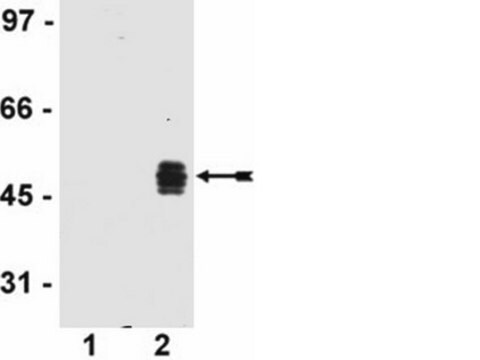
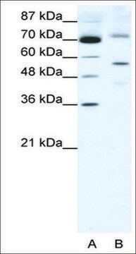
Western Blotting Analysis: A representative lot detected CD47 distribution among human polymorphonuclear (PMN) neutrophil subcellular fractions. Majority of CD47 was found co-localized with CD11b in the CD11b and secondary granules in unstimulated PMN and upregulated plasma membrane localization was seen following fMLP stimulation (Parkos, C.A., et al. (1996). J. Cell Biol. 132(3):437-450).
Research Category
Inflammation & Immunology
This mouse monoclonal Anti-CD47, clone PF3.1 Antibody, Cat. No. MABF958 is validated for use in Flow Cytometry, Immunoprecipitation, and Western Blotting for the detection of CD47.

Calidad

Evaluated by Flow Cytometry in human PBMCs.

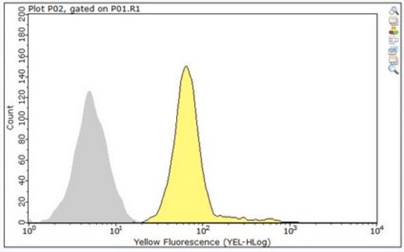
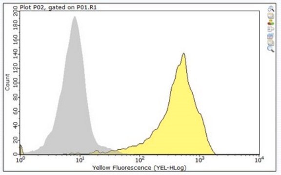
Flow Cytometry Analysis: 0.2 µL of this antibody detected CD47 surface expression on the gated lymphocytes population among one million human PBMCs.

Descripción de destino

33.47/30.00/31.45/32.10 kDa (mature isoform OA3-323/OA3-293/OA3-305/OA3-312) calculated. ~60-65 kDa (glycosylated) and ~35 kDa (deglycosylated) reported (Parkos, C.A., et al. (1996). J. Cell Biol. 132(3):437-450).

Forma física

Format: Purified
Protein G purified.
Purified mouse IgG1 in buffer containing 0.1 M Tris-Glycine (pH 7.4), 150 mM NaCl with 0.05% sodium azide.

Almacenamiento y estabilidad

Stable for 1 year at 2-8°C from date of receipt.

Otras notas

Concentration: Please refer to lot specific datasheet.

Cláusula de descargo de responsabilidad

Unless otherwise stated in our catalog or other company documentation accompanying the product(s), our products are intended for research use only and are not to be used for any other purpose, which includes but is not limited to, unauthorized commercial uses, in vitro diagnostic uses, ex vivo or in vivo therapeutic uses or any type of consumption or application to humans or animals.

¿No encuentra el producto adecuado?  

Pruebe nuestro Herramienta de selección de productos.

Código de clase de almacenamiento

12 - Non Combustible Liquids

Clase de riesgo para el agua (WGK)

WGK 1

Punto de inflamabilidad (°F)

Not applicable

Punto de inflamabilidad (°C)

Not applicable


Certificados de análisis (COA)

Busque Certificados de análisis (COA) introduciendo el número de lote del producto. Los números de lote se encuentran en la etiqueta del producto después de las palabras «Lot» o «Batch»

¿Ya tiene este producto?

Encuentre la documentación para los productos que ha comprado recientemente en la Biblioteca de documentos.

Visite la Librería de documentos

Nuestro equipo de científicos tiene experiencia en todas las áreas de investigación: Ciencias de la vida, Ciencia de los materiales, Síntesis química, Cromatografía, Analítica y muchas otras.

Póngase en contacto con el Servicio técnico