Direkt zum Inhalt
Merck
Alle Fotos(2)

Key Documents

MABT538

Sigma-Aldrich

Anti-Lamin A/C Antibody, clone 2E8.2

clone 2E8.2, from mouse

Synonym(e):

Prelamin-A/C, Lamin-A/C, Renal carcinoma antigen NY-REN-32

Anmeldenzur Ansicht organisationsspezifischer und vertraglich vereinbarter Preise


About This Item

UNSPSC-Code:
12352203
eCl@ss:
32160702

Biologische Quelle

mouse

Qualitätsniveau

Antikörperform

purified antibody

Antikörper-Produkttyp

primary antibodies

Klon

2E8.2, monoclonal

Speziesreaktivität

human

Methode(n)

immunohistochemistry: suitable (paraffin)
western blot: suitable

Isotyp

IgG1κ

NCBI-Hinterlegungsnummer

UniProt-Hinterlegungsnummer

Versandbedingung

ambient

Posttranslationale Modifikation Target

unmodified

Angaben zum Gen

human ... LMNA(4000)

Allgemeine Beschreibung

Lamin-A is a key component of the nuclear lamina, a protein network underlying the inner membrane that determines nuclear shape and size. The same gene (Lmna, Dhe, or Lamn1 in murine species; Gene ID 16905) that encodes lamin-A also codes for the alternatively spliced prelamin-C isoforms (UniProt P48678-2 and P48678-3). Murine lamin-A (UniProt P48678-1) is initially produced as a 665-amino acid prelamin A, which is further processed posttranslationally by four enzymatic steps to yield the mature Lamin-A. Farnesylation of Cys662, endoproteolytic cleavage of the last three amino acids (a.a. 663-665), carboxyl methylation of Cys662, and the final endoproteolytic removal of the remaining propeptide sequence (a.a. 648-662), including the farnesylated Cys662, catalyzed by the ER membrane zinc metalloproteinase ZMPSTE24.

Spezifität

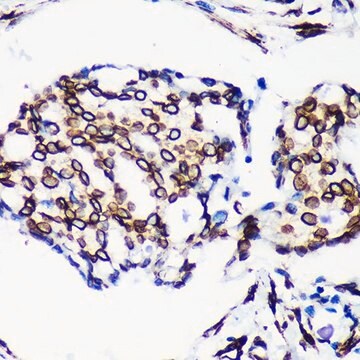
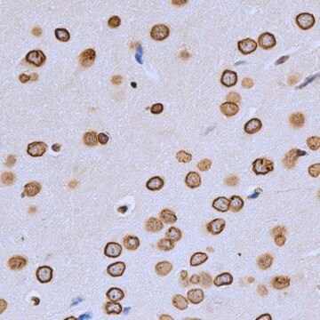
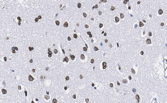
Clone 2E8.2 immunostained nuclear membrane in human tissue sections.

Immunogen

GST-tagged recombinant fragment within the C-terminal half of human Lamin A.

Anwendung

Research Category
Zellstruktur
Immunohistochemistry Analysis: A 1:1,000 dilution from a representative lot immunostained nuclear membrane in human cerebellum, kidney, and pancreas tissue sections.
This mouse monoclonal Anti-Lamin A/C Antibody, clone 2E8.2, Cat. No. MABT538 is validated for use in Immunohistochemistry (Paraffin) and Western Blotting for the detection of lamin A/C.

Qualität

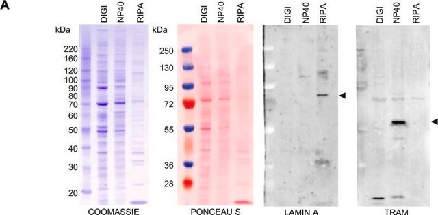
Evaluated by Western Blotting in A431 cell lysate.

Western Blotting Analysis: 0.5 µg/mL of this antibody detected lamin A/C in 10 µg of A431 cell lysate.

Zielbeschreibung

~70/65 kDa observed. 74.14/73.81/72.22 kDa (perpro-/pre-/mature Lamin A) and 65.14 kDa (Lamin C) calculated. Uncharacterized bands may be observed in some lysate(s).

Verlinkung

Replaces: MAB3211

Physikalische Form

Protein G purified.
Format: Purified
Purified mouse IgG1κ in buffer containing 0.1 M Tris-Glycine (pH 7.4), 150 mM NaCl with 0.05% sodium azide.

Lagerung und Haltbarkeit

Stable for 1 year at 2-8°C from date of receipt.

Sonstige Hinweise

Concentration: Please refer to lot specific datasheet.

Haftungsausschluss

Unless otherwise stated in our catalog or other company documentation accompanying the product(s), our products are intended for research use only and are not to be used for any other purpose, which includes but is not limited to, unauthorized commercial uses, in vitro diagnostic uses, ex vivo or in vivo therapeutic uses or any type of consumption or application to humans or animals.

Sie haben nicht das passende Produkt gefunden?  

Probieren Sie unser Produkt-Auswahlhilfe. aus.

Empfehlung

Lagerklassenschlüssel

12 - Non Combustible Liquids

WGK

WGK 1

Flammpunkt (°F)

Not applicable

Flammpunkt (°C)

Not applicable


Analysenzertifikate (COA)

Suchen Sie nach Analysenzertifikate (COA), indem Sie die Lot-/Chargennummer des Produkts eingeben. Lot- und Chargennummern sind auf dem Produktetikett hinter den Wörtern ‘Lot’ oder ‘Batch’ (Lot oder Charge) zu finden.

Besitzen Sie dieses Produkt bereits?

In der Dokumentenbibliothek finden Sie die Dokumentation zu den Produkten, die Sie kürzlich erworben haben.

Die Dokumentenbibliothek aufrufen

Zhuo Zhou et al.
Signal transduction and targeted therapy, 6(1), 382-382 (2021-11-05)
The global coronavirus disease 2019 (COVID-19) pandemic is caused by severe acute respiratory syndrome coronavirus 2 (SARS-CoV-2), a positive-sense RNA virus. How the host immune system senses and responds to SARS-CoV-2 infection remain largely unresolved. Here, we report that SARS-CoV-2

Unser Team von Wissenschaftlern verfügt über Erfahrung in allen Forschungsbereichen einschließlich Life Science, Materialwissenschaften, chemischer Synthese, Chromatographie, Analytik und vielen mehr..

Setzen Sie sich mit dem technischen Dienst in Verbindung.