Passa al contenuto
Merck
Tutte le immagini(1)

Documenti

131M-9

Sigma-Aldrich

CD31 (JC70) Mouse Monoclonal Antibody

Autenticatiper visualizzare i prezzi riservati alla tua organizzazione & contrattuali


About This Item

Codice UNSPSC:
12352203
NACRES:
NA.41

Origine biologica

mouse

Livello qualitativo

100
500

Coniugato

unconjugated

Forma dell’anticorpo

culture supernatant

Tipo di anticorpo

primary antibodies

Clone

JC70, monoclonal

Descrizione

For In Vitro Diagnostic Use in Select Regions (See Chart)

Forma fisica

buffered aqueous solution

Reattività contro le specie

human

Confezionamento

vial of 0.1 mL concentrate (131M-94)
vial of 0.5 mL concentrate (131M-95)
bottle of 1.0 mL predilute (131M-97)
vial of 1.0 mL concentrate (131M-96)
bottle of 7.0 mL predilute (131M-98)

Produttore/marchio commerciale

Cell Marque

tecniche

immunohistochemistry (formalin-fixed, paraffin-embedded sections): 1:25-1:100

Isotipo

IgG1κ

Controllo

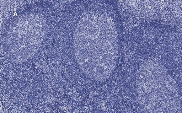
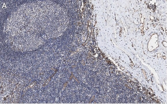
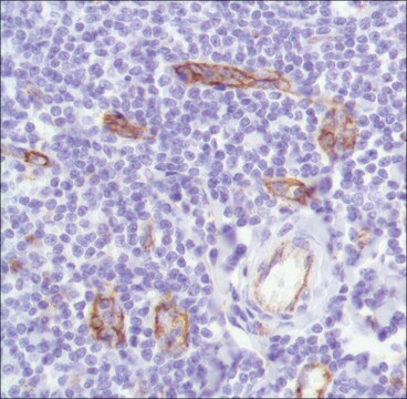
tonsil

Condizioni di spedizione

wet ice

Temperatura di conservazione

2-8°C

Visualizzazione

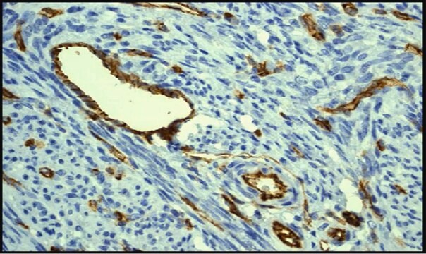
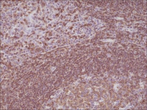
cytoplasmic, membranous

Informazioni sul gene

human ... PECAM1(5175)

Categorie correlate

Descrizione generale

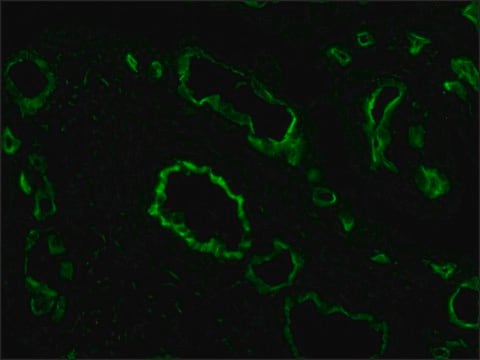
CD31, or platelet/endothelial cell adhesion molecule-1 (PECAM-1), is a vascular cell adhesionand signaling molecule that plays a role in maintaining the vascular permeability barrierat endothelial cell junctions. Immunohistochemical detection of CD31 shows cytoplasmicand membranous expression in non-neoplastic and neoplastic vascular endothelial cells,including neoplasms of vascular origin such as hemangiomas and angiosarcomas.

Linkage

CD31 Positive Control Slides, Product No. 131S, are available for immunohistochemistry (formalin-fixed, paraffin-embedded sections).

Stato fisico

Solution in Tris Buffer, pH 7.3-7.7, with 1% BSA and <0.1% Sodium Azide

Nota sulla preparazione

Download the IFU specific to your product lot and formatNote: This requires a keycode which can be found on your packaging or product label.

Altre note

For Technical Service please contact: 800-665-7284 or email: service@cellmarque.com

Note legali

Cell Marque is a trademark of Merck KGaA, Darmstadt, Germany

Not finding the right product?  

Try our Motore di ricerca dei prodotti.

Codice della classe di stoccaggio

12 - Non Combustible Liquids

Classe di pericolosità dell'acqua (WGK)

WGK 2

Punto d’infiammabilità (°F)

Not applicable

Punto d’infiammabilità (°C)

Not applicable


Certificati d'analisi (COA)

Cerca il Certificati d'analisi (COA) digitando il numero di lotto/batch corrispondente. I numeri di lotto o di batch sono stampati sull'etichetta dei prodotti dopo la parola ‘Lotto’ o ‘Batch’.

Possiedi già questo prodotto?

I documenti relativi ai prodotti acquistati recentemente sono disponibili nell’Archivio dei documenti.

Visita l’Archivio dei documenti

R L Attanoos et al.
Thorax, 55(10), 860-863 (2000-09-19)
Three cases of diffuse malignant vascular tumours of the pleura are described which mimicked malignant mesothelioma clinically and pathologically (so called "pseudomesothelioma"). All had occupational histories of exposure to asbestos. The relationship of these tumours to mesothelioma and asbestos exposure
Malak Abedalthagafi et al.
Annals of diagnostic pathology, 14(1), 15-22 (2010-02-04)
Cutaneous angiosarcoma (AS) is a rare malignant neoplasm of dermis composed of infiltrating cells of endothelial phenotype with overall poor prognosis. Although autocrine stimulation by vascular endothelial growth factor secretion may play a role in the pathogenesis of angiosarcoma, its
Xiu-Lian Xu et al.
The American Journal of dermatopathology, 32(8), 837-840 (2010-10-01)
Microvenular hemangioma (MVH) is an uncommon benign vascular neoplasm that usually occurs as a solitary asymptomatic red or purple papule, nodule, or plaque with a predilection for the upper extremities. Patients with more than 1 lesion, that is, multiple MVHs
CD31: An Immunospecific Marker for Endothelial Differentiation in Human Neoplasms
DeYoung BR, et al.
Appl. Immunohistochem., 1, 97-100 (1993)
F Alexander-Sefre et al.
Journal of clinical pathology, 56(10), 786-788 (2003-09-30)
Lymphovascular space invasion (LVSI) is an important step in the complex process of tumour metastasis. Various methods have been used in the past to improve the histological detection of LVSI. To develop a sensitive immunohistochemical method for the detection of

Il team dei nostri ricercatori vanta grande esperienza in tutte le aree della ricerca quali Life Science, scienza dei materiali, sintesi chimica, cromatografia, discipline analitiche, ecc..

Contatta l'Assistenza Tecnica.