Passa al contenuto
Merck
Tutte le immagini(2)

Documenti fondamentali

MABN737

Sigma-Aldrich

Anti-OPA1, clone 1OPA-1A8 Antibody

ascites fluid, clone 10PA-1A8, from mouse

Sinonimo/i:

Dynamin-like 120 kDa protein, mitochondrial, Large GTP-binding protein, LargeG, Optic atrophy protein 1 homolog, Dynamin-like 120 kDa protein, form S1

Autenticatiper visualizzare i prezzi riservati alla tua organizzazione & contrattuali


About This Item

Codice UNSPSC:
12352203
eCl@ss:
32160702
NACRES:
NA.41

Origine biologica

mouse

Livello qualitativo

Forma dell’anticorpo

ascites fluid

Tipo di anticorpo

primary antibodies

Clone

10PA-1A8, monoclonal

Reattività contro le specie

mouse, rat, human

tecniche

immunocytochemistry: suitable
immunoprecipitation (IP): suitable
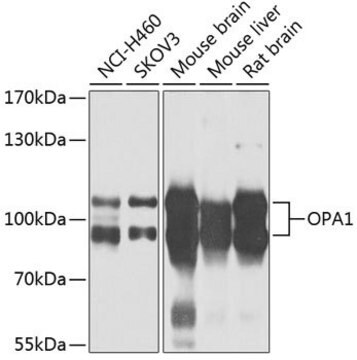
western blot: suitable

Isotipo

IgG1κ

N° accesso NCBI

N° accesso UniProt

Condizioni di spedizione

wet ice

modifica post-traduzionali bersaglio

unmodified

Informazioni sul gene

human ... OPA1(4976)

Descrizione generale

OPA1 (optic atrophy 1) is a dynamin-related GTPase that plays a role in genome preservation and mitochondrial morphology. Localization of OPA1 is thought to be anchored to the inner membrane of the mitochondria. OPA1 is highly expressed in inner and outer retinal neurons and is important for normal mitochondrial function in those cells. Mutations in OPA1 are the major cause of autosomal dominant optic atrophy. Recent research has found that OPA1 also has the ability to protect neurons from excitotoxic injury and could be a potential clinical target to increase endurance of neurons post brain damage.

Specificità

This antibody detects six known isoforms of OPA1 (Akepati, V. R., et al. (2008). J Neurochem. 106(1):372-383.).

Immunogeno

Recombinant protein corresponding to mouse OPA1.

Applicazioni

Immunocytochemistry Analysis: A 1:100 dilution from a representative lot detected OPA1 in COS cells (Prof. Mustapha Oulad-Abdelghani, IGBMC, France).

Immunoprecipitation Analysis: A representative lot from an independent laboratory immunoprecipitated OPA1 from mouse brain tissue lysate (Akepati, V. R., et al. (2008). J Neurochem. 106(1):372-383.).
Research Category
Neuroscience
Research Sub Category
Neurodegenerative Diseases
This Anti-OPA1 antibody, clone 1OPA-1A8 is validated for use in western blotting, ICC & IP for the detection of OPA1.

Qualità

Evaluated by human brain tissue lysate.

Western Blotting Analysis: A 1:1,000 dilution of this antibody detected OPA1 in 10 µg of human brain tissue lysate.

Descrizione del bersaglio

~90 and ~100 kDa observed. Six isoforms between 80 kDa and 100 kDa are known to exist (Akepati, V. R., et al. (2008). J Neurochem. 106(1):372-383.).

Stato fisico

Mouse monoclonal IgG1κ ascites containing 0.05% sodium azide.
Unpurified

Stoccaggio e stabilità

Stable for 1 year at -20°C from date of receipt.
Handling Recommendations: Upon receipt and prior to removing the cap, centrifuge the vial and gently mix the solution. Aliquot into microcentrifuge tubes and store at -20°C. Avoid repeated freeze/thaw cycles, which may damage IgG and affect product performance.

Esclusione di responsabilità

Unless otherwise stated in our catalog or other company documentation accompanying the product(s), our products are intended for research use only and are not to be used for any other purpose, which includes but is not limited to, unauthorized commercial uses, in vitro diagnostic uses, ex vivo or in vivo therapeutic uses or any type of consumption or application to humans or animals.

Non trovi il prodotto giusto?  

Prova il nostro Motore di ricerca dei prodotti.

Codice della classe di stoccaggio

12 - Non Combustible Liquids

Classe di pericolosità dell'acqua (WGK)

nwg

Punto d’infiammabilità (°F)

Not applicable

Punto d’infiammabilità (°C)

Not applicable


Certificati d'analisi (COA)

Cerca il Certificati d'analisi (COA) digitando il numero di lotto/batch corrispondente. I numeri di lotto o di batch sono stampati sull'etichetta dei prodotti dopo la parola ‘Lotto’ o ‘Batch’.

Possiedi già questo prodotto?

I documenti relativi ai prodotti acquistati recentemente sono disponibili nell’Archivio dei documenti.

Visita l’Archivio dei documenti

Vasudheva Reddy Akepati et al.
Journal of neurochemistry, 106(1), 372-383 (2008-04-19)
OPA1, a nuclear encoded mitochondrial protein causing autosomal dominant optic atrophy, is a key player in mitochondrial fusion and cristae morphology regulation. In the present study, we have compared the OPA1 transcription and translation products of different mouse tissues. Unlike

Il team dei nostri ricercatori vanta grande esperienza in tutte le aree della ricerca quali Life Science, scienza dei materiali, sintesi chimica, cromatografia, discipline analitiche, ecc..

Contatta l'Assistenza Tecnica.