Accéder au contenu
Merck
Toutes les photos(1)

Principaux documents

335M-9

Sigma-Aldrich

Cytokeratin (35betaH11) Mouse Monoclonal Antibody

Se connecterpour consulter vos tarifs contractuels et ceux de votre entreprise/organisme


About This Item

Code UNSPSC :
12352203
Nomenclature NACRES :
NA.41

Source biologique

mouse

Niveau de qualité

100
500

Conjugué

unconjugated

Forme d'anticorps

culture supernatant

Type de produit anticorps

primary antibodies

Clone

35betaH11, monoclonal

Description

For In Vitro Diagnostic Use in Select Regions (See Chart)

Forme

buffered aqueous solution

Espèces réactives

human

Conditionnement

vial of 0.1 mL concentrate (335M-94)
vial of 0.5 mL concentrate (335M-95)
bottle of 1.0 mL predilute (335M-97)
vial of 1.0 mL concentrate (335M-96)
bottle of 7.0 mL predilute (335M-98)

Fabricant/nom de marque

Cell Marque

Technique(s)

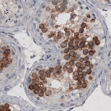
immunohistochemistry (formalin-fixed, paraffin-embedded sections): 1:100-1:500

Isotype

IgMκ

Contrôle

prostate

Conditions d'expédition

wet ice

Température de stockage

2-8°C

Visualisation

cytoplasmic

Informations sur le gène

human ... KRT1(3848)

Description générale

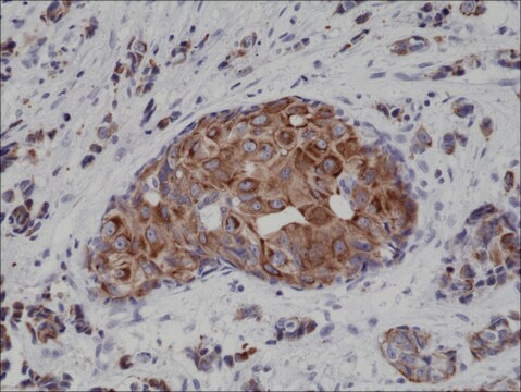
Anti-Cytokeratin 8 stains most non-squamous epithelial tumors, squamous tumors are negative for this antibody as a rule. This antibody stains adenocarcinomas of the breast, ovary, gastrointestinal tract, thyroid, pancreas, bile duct, and salivary glands. This antibody does not react with skeletal muscle or nerve cells.

Qualité


IVD

IVD

IVD

RUO

Liaison

Cytokeratin (35betaH11) Positive Control Slides, Product No. 335S, are available for immunohistochemistry (formalin-fixed, paraffin-embedded sections).

Forme physique

Solution in Tris Buffer, pH 7.3-7.7, with 1% BSA and <0.1% Sodium Azide

Notes préparatoires

Download the IFU specific to your product lot and formatNote: This requires a keycode which can be found on your packaging or product label.

Autres remarques

For Technical Service please contact: 800-665-7284 or email: service@cellmarque.com

Informations légales

Cell Marque is a trademark of Merck KGaA, Darmstadt, Germany

Vous ne trouvez pas le bon produit ?  

Essayez notre Outil de sélection de produits.


Faites votre choix parmi les versions les plus récentes :

Certificats d'analyse (COA)

Lot/Batch Number

Désolés, nous n'avons pas de COA pour ce produit disponible en ligne pour le moment.

Si vous avez besoin d'assistance, veuillez contacter Service Clients

Déjà en possession de ce produit ?

Retrouvez la documentation relative aux produits que vous avez récemment achetés dans la Bibliothèque de documents.

Consulter la Bibliothèque de documents

A C Knapp et al.
Cell, 59(1), 67-79 (1989-10-06)
Intermediate filaments (IFs) of the cytokeratin (CK) type are cytoskeletal elements typical for epithelial differentiation. However, in diverse transformed culture lines of non-epithelial origin, rare cells emerge spontaneously, which synthesize, in addition to their vimentin IFs, CKs 8 and 18.
R Eichner et al.
The Journal of cell biology, 98(4), 1388-1396 (1984-04-01)
Human epidermal keratinocytes express under various growth conditions a total of at least nine keratins that can be divided into two subfamilies. Subfamily A comprises 40-, 46-, 48-, 50-/50'-, and 56.5-kilodalton (kd) keratins which are relatively acidic (pI less than
A M Gown et al.
American journal of clinical pathology, 84(4), 413-424 (1985-10-01)
A panel of monoclonal antibodies to human intermediate filament proteins was tested on an unselected series of 246 neoplasms. The antibody panel includes two different anti-cytokeratin antibodies, an anti-vimentin antibody, and an anti-neurofilament antibody (Gown and Vogel, Am J Pathol
T T Sun et al.
The Journal of investigative dermatology, 81(1 Suppl), 109s-115s (1983-07-01)
Keratins are a group of water-insoluble proteins (molecular weight range 40-70 K) that form 10-nm tonofilaments in a wide variety of epithelial cells. The subunit composition of the keratin filaments varies with cell type, period of embryonic development, stage of
H Battifora
The American journal of surgical pathology, 12 Suppl 1, 24-42 (1988-01-01)
The past few years have seen great progress in our knowledge of the biology and the chemistry of intermediate filaments and other filamentous proteins. This has translated into the development of antibodies that, together with new, highly sensitive immunohistochemical methods

Notre équipe de scientifiques dispose d'une expérience dans tous les secteurs de la recherche, notamment en sciences de la vie, science des matériaux, synthèse chimique, chromatographie, analyse et dans de nombreux autres domaines..

Contacter notre Service technique