MAB1569
Anti-Microglia Antibody, clone 25F9
clone 25F9, Chemicon®, from mouse
Synonyme(s) :
Macrophages, Late State Inflammatory
About This Item
Produits recommandés
Source biologique
mouse
Niveau de qualité
Forme d'anticorps
purified immunoglobulin
Type de produit anticorps
primary antibodies
Clone
25F9, monoclonal
Espèces réactives
human
Fabricant/nom de marque
Chemicon®
Technique(s)
flow cytometry: suitable
immunocytochemistry: suitable
immunohistochemistry: suitable
Isotype
IgG1
Conditions d'expédition
wet ice
Modification post-traductionnelle de la cible
unmodified
Description générale
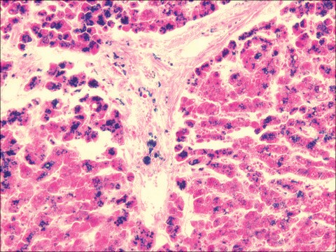
Isolated cells: Absent on freshly isolated monocytes and other blood cells; present on 40-50% of human monocytes after 6-7 day culture, also positive on some melanoma and carcinoma cell lines. Tissue sections: Kupffer cells, histiocytes (skin), macrophages of the thymus, in the germinal centers of lymph nodes and spleen, in mamma carcinoma, melanoma, osteocarcinoma and gastric cancer; excema, sarcoidosis, BCG granuloma; synovial lining cells, tuberculoid leprosy; no expression in lepramatous leprosy.
Spécificité
Antigen distribution: absent from freshly isolated monocytes and other blood cells; present on 40-50% of human monocytes after 6-7 days in culture, also positive on some melanoma and carcinoma lines. In tissue sections, clone identifies Kupffer cells, histiocytes (skin), macrophages of the thymus, in te germinal centers of lymph nodes and spleen, in mamma carcinoma, melanoma, osteocarcinoma and gastric cancer; excema, sarcoidosis, BCG granuloma;synovial lining cells, tuberculoid leprosy, however no expression in lepramatous leprosy.
Species reactivity is seen in human, rhesus monkey, and pig, other species not tested.
Application
Immunocytochemistry
Optimal working dilutions must be determined by the end user.
Forme physique
Stockage et stabilité
Informations légales
Vous ne trouvez pas le bon produit ?
Essayez notre Outil de sélection de produits.
Mention d'avertissement
Warning
Mentions de danger
Conseils de prudence
Classification des risques
Acute Tox. 4 Dermal - Acute Tox. 4 Inhalation - Aquatic Chronic 3
Code de la classe de stockage
11 - Combustible Solids
Classe de danger pour l'eau (WGK)
WGK 3
Certificats d'analyse (COA)
Recherchez un Certificats d'analyse (COA) en saisissant le numéro de lot du produit. Les numéros de lot figurent sur l'étiquette du produit après les mots "Lot" ou "Batch".
Déjà en possession de ce produit ?
Retrouvez la documentation relative aux produits que vous avez récemment achetés dans la Bibliothèque de documents.
Notre équipe de scientifiques dispose d'une expérience dans tous les secteurs de la recherche, notamment en sciences de la vie, science des matériaux, synthèse chimique, chromatographie, analyse et dans de nombreux autres domaines..
Contacter notre Service technique


