Skip to Content
Merck
All Photos(1)

Key Documents

C3556

Sigma-Aldrich

Catalase from human erythrocytes

≥90% (SDS-PAGE), buffered aqueous solution, ≥30,000 units/mg protein

Synonym(s):

H2O2:H2O2 oxidoreductase

Sign Into View Organizational & Contract Pricing


About This Item

CAS Number:
Enzyme Commission number:
EC Number:
MDL number:
UNSPSC Code:
12352204
NACRES:
NA.54

biological source

human erythrocytes

Quality Level

Assay

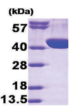
≥90% (SDS-PAGE)

form

buffered aqueous solution

specific activity

≥30,000 units/mg protein

mol wt

tetramer ~250 kDa

concentration

≤10 mg/mL

technique(s)

cell based assay: suitable

UniProt accession no.

shipped in

wet ice

storage temp.

−20°C

InChI

1S/C9H10O3/c1-2-12-9(11)7-3-5-8(10)6-4-7/h3-6,10H,2H2,1H3

InChI key

NUVBSKCKDOMJSU-UHFFFAOYSA-N

Gene Information

human ... CAT(847)

General description

Research area: Cell Signaling

Human catalase is a member of the monofunctional heme-containing catalases. It is an intracellular enzyme located at higher concentrations in the liver, erythrocytes, and kidney. Catalase is a homo-tetrameric protein and comprises amino acid residues, one heme group that is iron III protoporphyrin IX, and a nicotinamide adenine dinucleotide phosphate (NADPH) molecule. It is a ubiquitous enzyme found in most aerobic organisms. The catalase (CAT) gene is located on the human chromosome at 11p13.

Application

Catalase from human erythrocytes has been used:

  • to prevent reoxidation of reduced cytochrome c by H2O2 while measuring the production of superoxide with cytochrome C.
  • as a component of the imaging buffer for stochastic optical reconstruction microscopy (STORM) imaging of human skin fibroblasts
  • as a component of the gloxy mix for single-molecule imaging

Biochem/physiol Actions

Deficiency of catalase and oxidative stress are associated with vitiligo, hypertension, Alzheimer′s disease, diabetes mellitus, and acatalasemia. Catalase shows a dichotomous role in cancer as its expression is at a higher level in chronic lymphocytic leukemia, glioma, melanoma, and gastric cancer and lower levels of expression are seen in acute myeloid leukemia, pancreatic, lung, prostate, and skin (non-melanoma) cancers.
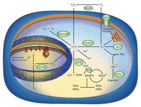
Catalase activates the decomposition of hydrogen peroxide, a reactive oxygen species, into water and oxygen. It functions as a natural antioxidant, protecting cells against oxidative damage to proteins, lipids and nucleic acids. Catalase has also been used to study the role reactive oxygen species play in gene expression and apoptosis.

Unit Definition

One unit will decompose 1.0 μmole of H2O2 per min at pH 7.0 at 25 °C, while the H2O2 conc. falls from 10.3 to 9.2 mM, measured by the rate of decrease of A240.

Physical form

Solution in 50 mM Tris, pH 8.0

Storage and Stability

Tightly closed. Dry. Keep locked up or in an area accessible only to qualified or authorized
persons

Analysis Note

Protein determined by biuret

inhibitor

Product No.
Description
Pricing

Pictograms

Health hazard

Signal Word

Danger

Hazard Statements

Precautionary Statements

Hazard Classifications

Resp. Sens. 1

Storage Class Code

11 - Combustible Solids

WGK

WGK 1

Flash Point(F)

Not applicable

Flash Point(C)

Not applicable

Personal Protective Equipment

dust mask type N95 (US), Eyeshields, Gloves

Choose from one of the most recent versions:

Certificates of Analysis (COA)

Lot/Batch Number

Don't see the Right Version?

If you require a particular version, you can look up a specific certificate by the Lot or Batch number.

Already Own This Product?

Find documentation for the products that you have recently purchased in the Document Library.

Visit the Document Library

Urte Neniskyte et al.
Methods in molecular biology (Clifton, N.J.), 1041, 103-111 (2013-07-03)
Reactive oxygen and nitrogen species are both regulators and effectors of microglial activation, and assays of these oxidants can be used as a measure of acute and chronic activation of microglial cells. Here we describe quick methods to assess the
Roshan L Shrestha et al.
Nature communications, 8(1), 150-150 (2017-07-29)
Human chromosomes are captured along microtubule walls (lateral attachment) and then tethered to microtubule-ends (end-on attachment) through a multi-step end-on conversion process. Upstream regulators that orchestrate this remarkable change in the plane of kinetochore-microtubule attachment in human cells are not
Bailin Zhao et al.
Nucleic acids research, 48(7), 3605-3618 (2020-02-14)
During non-homologous DNA end joining (NHEJ), bringing two broken dsDNA ends into proximity is an essential prerequisite for ligation by XRCC4:Ligase IV (X4L4). This physical juxtaposition of DNA ends is called NHEJ synapsis. In addition to the key NHEJ synapsis proteins
Hui-Min Yin et al.
Science advances, 6(19), eaay9466-eaay9466 (2020-06-05)
The cardiac and hematopoietic progenitors (CPs and HPs, respectively) in the mesoderm ultimately form a well-organized circulation system, but mechanisms that reconcile their development remain elusive. We found that activating transcription factor 3 (atf3) was highly expressed in the CPs
Junwei Li et al.
Science translational medicine, 12(558) (2020-08-28)
Epithelial tissues line the organs of the body, providing an initial protective barrier as well as a surface for nutrient and drug absorption. Here, we identified enzymatic components present in the gastrointestinal epithelium that can serve as selective means for

Articles

Oxidative stress is mediated, in part, by reactive oxygen species produced by multiple cellular processes and controlled by cellular antioxidant mechanisms such as enzymatic scavengers or antioxidant modulators. Free radicals, such as reactive oxygen species, cause cellular damage via cellular.

Our team of scientists has experience in all areas of research including Life Science, Material Science, Chemical Synthesis, Chromatography, Analytical and many others.

Contact Technical Service