Direkt zum Inhalt
Merck
Alle Fotos(1)

Wichtige Dokumente

NE1017

Sigma-Aldrich

Anti-Pan-Neuronal Neurofilament Marker Mouse mAb (SMI-311)

liquid, clone SMI-311, Calbiochem®

Anmeldenzur Ansicht organisationsspezifischer und vertraglich vereinbarter Preise


About This Item

UNSPSC-Code:
12352203

Biologische Quelle

mouse

Qualitätsniveau

Antikörperform

ascites fluid

Antikörper-Produkttyp

primary antibodies

Klon

SMI-311, monoclonal

Form

liquid

Enthält

≤0.1% sodium azide as preservative

Speziesreaktivität (Voraussage durch Homologie)

mammals

Hersteller/Markenname

Calbiochem®

Lagerbedingungen

OK to freeze
avoid repeated freeze/thaw cycles

Isotyp

IgG1, IgGM

Versandbedingung

wet ice

Lagertemp.

−20°C

Posttranslationale Modifikation Target

unmodified

Angaben zum Gen

human ... NEFL(4747)

Allgemeine Beschreibung

Mouse monoclonal antibody supplied as undiluted ascites. Recognizes the ~180-200 kDa pan-neuronal neurofilament marker protein.
Recognizes ~180-200 kDa pan-neuronal neurofilament marker in rat central nervous system cytoskeletal preparations.
This Anti-Pan-Neuronal Neurofilament Marker Mouse mAb (SMI-311) is validated for use in ELISA, Frozen Sections, WB, ICC, Paraffin Sections for the detection of Pan-Neuronal Neurofilament Marker.

Immunogen

Rat
homogenized hypothalami extracted from Fischer 344 rat brain

Anwendung


ELISA (1:1000)
Frozen Sections (1:1000, see comments)
Immunoblotting (1:1000)
Immunocytochemistry (1:1000, see comments)
Paraffin Sections (1:1000, heat pre-treatment required, see comments)

Warnhinweis

Toxicity: Standard Handling (A)

Physikalische Form

Undiluted ascites.

Rekonstituierung

Following initial thaw, aliquot and freeze (-20°C).

Hinweis zur Analyse

Positive Control
Rat brain or rat CNS cytoskeletal preparations

Sonstige Hinweise

Agostino, A., et al. 2003. Hum. Mol. Genet.12, 399.
Tsunoda, I., et al. 2003. Am. J. Pathol.162, 1259.
Ulfig, N., et al. 1998. Cell Tissue Res.291, 433.
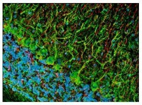
This antibody cocktail was selected to provide a specific marker for neurons in tissue sections and cultured cells. In contrast to individual anti-nonphosphoneurofilament antibodies that identify different subsets of neurons, this cocktail is a convenient marker for neurons in general and their differentiation from non-neuronal cells. Also useful as an early marker of neuronal migration and differentiation in human fetal development, yielding Golgi-like images without the disadvantages of the lack of selectivity and poor specificity of the Golgi technique. Can be used to trace the "inside-out gradient" of neuron production and differentiation in specifically delineating cell bodies and dendrites. Certain pathologic conditions, such as malnutrition, affect the SMI-311-visualized soma size and dendritic arborization. Tissues and cultured cells can be fixed in a variety of paraformaldehyde- or formaldehyde-containing fixatives, including Bouin′s fixative. This antibody does not react well with tissues or cells fixed in glutaraldehyde/paraformaldehyde. For staining paraffin sections it is recommended that de-paraffinized tissues be autoclaved in dH2O for 10 min to expose the epitope. Trypsin pre-treatment will abolish reactivity. Post-fixation in cold methanol or methanol/hydrogen peroxide facilitates access of the antibody to the neurons in frozen sections and thick tissues sections that have been fixed in 4% paraformaldehyde or cultured cells. For tissues that have not been paraffin-embedded and have been stored for long periods of time in formaldehyde fixatives, it is recommended that the tissues (up to 0.5 cm thick) be boiled in Tris-buffered saline, pH 9.0 for 15 min prior to sectioning. Antibody should be titrated for optimal results in individual systems.

Rechtliche Hinweise

CALBIOCHEM is a registered trademark of Merck KGaA, Darmstadt, Germany

Sie haben nicht das passende Produkt gefunden?  

Probieren Sie unser Produkt-Auswahlhilfe. aus.

Lagerklassenschlüssel

10 - Combustible liquids

WGK

WGK 1


Analysenzertifikate (COA)

Suchen Sie nach Analysenzertifikate (COA), indem Sie die Lot-/Chargennummer des Produkts eingeben. Lot- und Chargennummern sind auf dem Produktetikett hinter den Wörtern ‘Lot’ oder ‘Batch’ (Lot oder Charge) zu finden.

Besitzen Sie dieses Produkt bereits?

In der Dokumentenbibliothek finden Sie die Dokumentation zu den Produkten, die Sie kürzlich erworben haben.

Die Dokumentenbibliothek aufrufen

Timo Schomann et al.
Cell and tissue research, 381(1), 55-69 (2020-02-10)
Traumatic brain injury (TBI) is a devastating event for which current therapies are limited. Stem cell transplantation may lead to recovery of function via different mechanisms, such as cell replacement through differentiation, stimulation of angiogenesis and support to the microenvironment.
Yue Zhang et al.
The Journal of biological chemistry, 292(1), 100-111 (2016-11-30)
Astrocytes respond to CNS insults through reactive astrogliosis, but the underlying mechanisms are unclear. In this study, we show that inactivation of mechanistic target of rapamycin complex (mTORC1) signaling in postnatal neurons induces reactive astrogliosis in mice. Ablation of Raptor
Hyun-Jeong Yang et al.
Nature communications, 7, 10884-10884 (2016-03-11)
While the formation of myelin by oligodendrocytes is critical for the function of the central nervous system, the molecular mechanism controlling oligodendrocyte differentiation remains largely unknown. Here we identify G protein-coupled receptor 37 (GPR37) as an inhibitor of late-stage oligodendrocyte

Unser Team von Wissenschaftlern verfügt über Erfahrung in allen Forschungsbereichen einschließlich Life Science, Materialwissenschaften, chemischer Synthese, Chromatographie, Analytik und vielen mehr..

Setzen Sie sich mit dem technischen Dienst in Verbindung.