Direkt zum Inhalt
Merck
Alle Fotos(3)

Key Documents

MABN1724

Sigma-Aldrich

Anti-GABA(A) Receptor α2 Antibody, clone N399/19

clone N399/19, from mouse

Synonym(e):

Gamma-aminobutyric acid receptor subunit alpha-2, GABA(A) receptor subunit alpha-2, Gamma-aminobutyric acid receptor A2 subunit

Anmeldenzur Ansicht organisationsspezifischer und vertraglich vereinbarter Preise


About This Item

UNSPSC-Code:
12352203
eCl@ss:
32160702
NACRES:
NA.41

Biologische Quelle

mouse

Qualitätsniveau

Antikörperform

purified immunoglobulin

Antikörper-Produkttyp

primary antibodies

Klon

N399/19, monoclonal

Speziesreaktivität

rat, human, mouse

Methode(n)

immunohistochemistry: suitable
western blot: suitable

Isotyp

IgG1κ

UniProt-Hinterlegungsnummer

Versandbedingung

wet ice

Posttranslationale Modifikation Target

unmodified

Angaben zum Gen

human ... GABRA2 (2555)

Allgemeine Beschreibung

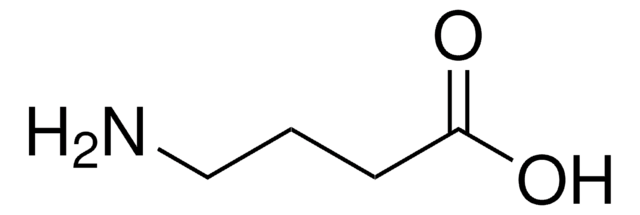
Gamma-aminobutyric acid receptor subunit alpha-2 (UniProt P23576; also known as GABA(A) receptor subunit alpha-2, Gamma-aminobutyric acid receptor A2 subunit) is encoded by the Gabra2 (also known as Gabra-2) gene (Gene ID 289606) in rat species. The GABAA receptor (GABAAR) is an ionotropic receptor and ligand-gated ion channel. Its endogenous ligand is the inhibitory neurotransmitter γ-aminobutyric acid (GABA) in the central nervous system (CNS). Upon activation, the GABAA receptor selectively conducts chloride ion through its pore, resulting in hyperpolarization of the neuron. The receptor is a pentameric complex composed of two alpha, two beta, and one gamma subunits arranged around a central pore. Each subunit contains four transmembrane domains with both the N- and C-terminus located extracellularly. In human and rat, there exist six types of alpha subunits (encoded by GABRA1-6), three types of beta subunits (encoded by GABRB1-3), and three types of gamma subunits (encoded by GABRG1-3). The alpha 2 subunit is intially produced with a signal peptide sequence (aa 1-28), which is then cleaved off to yield the mature subunit (aa 29-451) with a large N-terminal extracellular domain (a.a. 29-251), four transmembrane segments (a.a. 252-273, 279-300, 313-334, 420-441) and a large intracellular/cytoplasmic loop (a.a. 335-419) between the third and fourth transmembrane domain.

Spezifität

Clone N399/19 detects the GABA(A) receptor subunit alpha-2 band only in brain membrane extracts from wild-type rats and mice, but not from Gabra2-knockout mice. Clone N399/19 does not cross-react with GABA(A) receptor subunit alpha-1.

Immunogen

Epitope: Cytoplasmic domain.
Recombinant rat GABA(A) receptor α2 cytoplasmic fragment.

Anwendung

Research Category
Neurowissenschaft
Research Sub Category
Ionenkanäle & -transporter
Detect GABA(A) Receptor α2 using this Anti-GABA(A) Receptor α2 Antibody, clone N399/19 validated for use in Western Blotting, Immunohistochemistry.
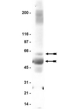
Western Blotting Analysis: 2.0 µg/mL from a representative lot detected GABA(A) receptor subunit alpha-2 in 10 µg rat brain tissue lysate.
Western Blotting Analysis: A representative lot detected the GABA(A) receptor subunit alpha-2 target band only in brain membrane extracts from wild-type rats and mice, but not from Gabra2-knockout mice.
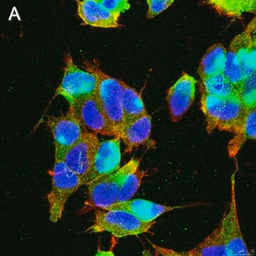
Immunohistochemisry Analysis: A representative lot detected GABA(A) receptor subunit alpha-2 immunoreactive regions, includinghippocampus, in rat whole brain sections (Courtesy of Professor. James S. Trimmer, UC Davis, CA, USA).

Qualität

Evaluated by Western Blotting in human brain tissue lysate.

Western Blotting Analysis: 2.0 µg/mL of this antibody detected GABA(A) receptor subunit alpha-2 in 10 µg human brain tissue lysate.

Zielbeschreibung

~57 kDa observed. Target band appears larger than the calculated molecular weight of 47.94 kDa due to glycosylation.

Physikalische Form

Protein G Purified
Format: Purified
Purified mouse monoclonal IgG1κ antibody in buffer containing 0.1 M Tris-Glycine (pH 7.4), 150 mM NaCl with 0.05% sodium azide.

Lagerung und Haltbarkeit

Stable for 1 year at 2-8°C from date of receipt.

Sonstige Hinweise

Concentration: Please refer to lot specific datasheet.

Haftungsausschluss

Unless otherwise stated in our catalog or other company documentation accompanying the product(s), our products are intended for research use only and are not to be used for any other purpose, which includes but is not limited to, unauthorized commercial uses, in vitro diagnostic uses, ex vivo or in vivo therapeutic uses or any type of consumption or application to humans or animals.

Sie haben nicht das passende Produkt gefunden?  

Probieren Sie unser Produkt-Auswahlhilfe. aus.

Lagerklassenschlüssel

12 - Non Combustible Liquids

WGK

WGK 1

Flammpunkt (°F)

Not applicable

Flammpunkt (°C)

Not applicable


Analysenzertifikate (COA)

Suchen Sie nach Analysenzertifikate (COA), indem Sie die Lot-/Chargennummer des Produkts eingeben. Lot- und Chargennummern sind auf dem Produktetikett hinter den Wörtern ‘Lot’ oder ‘Batch’ (Lot oder Charge) zu finden.

Besitzen Sie dieses Produkt bereits?

In der Dokumentenbibliothek finden Sie die Dokumentation zu den Produkten, die Sie kürzlich erworben haben.

Die Dokumentenbibliothek aufrufen

Pradeep Kannampalli et al.
Neuroscience, 346, 349-364 (2017-01-28)
Painful events early in life have been shown to increase the incidence of interstitial cystitis/painful bladder syndrome in adulthood. However, the intrinsic mechanism is not well studied. We previously reported that neonatal bladder inflammation causes chronic visceral hypersensitivity along with
Gloria S Forkuo et al.
Molecular pharmaceutics, 13(6), 2026-2038 (2016-04-28)
Recent studies have demonstrated that subtype-selective GABAA receptor modulators are able to relax precontracted human airway smooth muscle ex vivo and reduce airway hyper-responsiveness in mice upon aerosol administration. Our goal in this study was to investigate systemic administration of

Unser Team von Wissenschaftlern verfügt über Erfahrung in allen Forschungsbereichen einschließlich Life Science, Materialwissenschaften, chemischer Synthese, Chromatographie, Analytik und vielen mehr..

Setzen Sie sich mit dem technischen Dienst in Verbindung.