S7101
Kit de détection in situ de l'apoptose ApopTag Plus Peroxydase
The ApopTag Plus Peroxidase In Situ Apoptosis Detection Kit detects apoptotic cells by labeling & detecting DNA strand breaks by the indirect TUNEL method.
Synonyme(s) :
Kit de détection de l'apoptose in situ
About This Item
Produits recommandés
Niveau de qualité
Fabricant/nom de marque
ApopTag
Chemicon®
Méthode de détection
colorimetric
Conditions d'expédition
dry ice
Description générale
Les kits ApopTag Peroxydase ont été validés pour une utilisation dans la coloration histochimique et cytochimique des échantillons suivants : tissus fixés au formaldéhyde et inclus en paraffine, coupes de cryostat, suspensions cellulaires, échantillons obtenus par Cytospin, et cultures cellulaires. Des méthodes de montage complètes ont été développées (34, 45). (Voir la section V. de la fiche technique : Références).
La spécificité de coloration des kits ApopTag Peroxydase a été démontrée par Chemicon et de nombreux autres laboratoires. Chemicon a testé de nombreux types de systèmes modèles cellulaires et tissulaires, notamment : (a) prostate, thymus et gros intestin humains (données internes) ; (b) prostate ventrale de rat post-castration (21), (c) lymphocytes de thymus de rat traités in vitro avec de la dexaméthasone (3, 13), (d) membres d'embryons de souris de 14 jours (1) et (e) glande mammaire de rat en régression après sevrage (36). Dans les modèles de thymocytes et de prostate, l'électrophorèse sur gel d'agarose a été utilisée pour évaluer la quantité d'ADN présentant une distribution "en échelle" (ou "DNA laddering" en anglais), dont le pic coïncide avec le pourcentage maximal de cellules colorées. De nombreux articles de revues scientifiques, publiés par des laboratoires du monde entier, ont établi l'utilité des kits ApopTag. (Voir la section V. de la fiche technique : Références, Publications citant les kits ApopTag).
Application
ApopTag In Situ Apoptosis Detection Kits label apoptotic cells in research samples by modifying DNA fragments utilizing terminal deoxynucleotidyl transferase (TdT) for detection of apoptotic cells by specific staining.
This manual contains information and protocols for the ApopTag Plus In Situ Apoptosis Detection Kit.
Principles of the Procedure
The reagents provided in ApopTag Peroxidase Kits are designed to label the free 3′OH DNA termini in situ with chemically labeled and unlabeled nucleotides. The nucleotides contained in the Reaction Buffer are enzymatically added to the DNA by terminal deoxynucleotidyl transferase (TdT) (13, 31). TdT catalyzes a template-independent addition of nucleotide triphosphates to the 3′-OH ends of double-stranded or single-stranded DNA. The incorporated nucleotides form an oligomer composed of digoxigenin-conjugated nucleotide and unlabeled nucleotide in a random sequence. The ratio of labeled to unlabeled nucleotide in ApopTag Peroxidase Kits is optimized to promote anti-digoxigenin antibody binding. The exact length of the oligomer added has not been measured.
DNA fragments which have been labeled with the digoxigenin-nucleotide are then allowed to bind an anti-digoxigenin antibody that is conjugated to a peroxidase reporter molecule (Figure 1A). The bound peroxidase antibody conjugate enzymatically generates a permanent, intense, localized stain from chromogenic substrates, providing sensitive detection in immunohistochemistry or immunocytochemistry (i.e. on tissue or cells). This mixed molecular biological-histochemical systems allows for sensitive and specific staining of very high concentrations of 3′-OH ends that are localized in apoptotic bodies.
The ApopTag system differs significantly from previously described in situ labeling techniques for apoptosis (13, 16, 38, 46), in which avidin binding to cellular biotin can be a source of error. The digoxigenin/anti-digoxigenin system has been found to be equally sensitive to avidin/biotin systems (22). The sole natural source of digoxigenin is the digitalis plant. Immunochemically-similar ligands for binding of the anti-digoxigenin antibody are generally insignificant in animal tissues, ensuring low background staining. Affinity purified sheep polyclonal antibody is the specific anti-digoxigenin reagent used in ApopTag Kits. This antibody exhibits <1% cross-reactivity with the major vertebrate steroids. In addition, the Fc portion of this antibody has been removed by proteolytic digestion to eliminate any non-specific adsorption to cellular Fc receptors.
Results using ApopTag Kits have been widely published (see Sec. V. References, Publications Citing ApopTag Kits). The ApopTag product line provides various options in experimental design. A researcher can choose to detect staining by brightfield or fluorescence microscopy or by flow cytometry, depending on available expertise and equipment. There are also opportunities to study other proteins of interest in the context of apoptosis when using ApopTag Kits. By using antibodies conjugated with an enzyme other than peroxidase and an appropriate choice of substrate, it is possible to simultaneously examine another protein and apoptosis using ApopTag Peroxidase Kits.
Composants
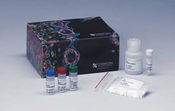
Tampon de réaction 91417 2,0 mL -15 °C à -25 °C
Enzyme TdT 90418 0,672 mL -15 °C à -25 °C
Tampon de lavage/arrêt 90419 20 mL -15 °C à -25 °C
Anticorps anti-digoxygénine conjugué à la peroxidase* 90420 3,0 mL 2 °C à 8 °C
Lamelles en plastique 90421 100 unités. Temp. ambiante
Lames de contrôle 90422 2 unités. Temp. ambiante
Substrat DAB 90423 130 µL 2 °C à 8 °C
Tampon de dilution DAB 90424 6,5 mL 2 °C à 8 °C
*Anticorps polyclonal de mouton purifié par affinité
Nombre d'échantillons par kit : Le kit fournit le matériel suffisant pour colorer 40 échantillons de tissu d'environ 5 cm2 chacun, lorsqu'il est utilisé conformément aux instructions. Si le kit est utilisé pour l'analyse d'échantillons sur lame, le tampon de réaction sera épuisé avant les autres réactifs.
Stockage et stabilité
Précautions
1. Les composants suivants du kit contiennent du cacodylate de potassium (acide diméthylarsinique) comme tampon : Tampon d'équilibration (90416), tampon de réaction (90417) et enzyme TdT (90418). Ces éléments sont nocifs en cas d'ingestion ; éviter tout contact avec la peau et les yeux (porter des gants et des lunettes) et rincer immédiatement en cas de contact.
2. Il a été démontré que le substrat DAB (3,3'-diaminobenzidine) (90423) est un cancérogène potentiel et que le contact avec la peau doit être évité. En cas de contact avec la peau, rincez abondamment à l'eau distillée (dH2O).
3. Les anticorps conjugués (90420) contiennent 0,08 % d'azide de sodium comme conservateur.
4. L'enzyme TdT (90418) contient du glycérol ; elle ne gèle pas à -20 °C. Pour une durée de conservation maximale, ne pas réchauffer ce réactif à température ambiante avant utilisation.
Informations légales
Clause de non-responsabilité
Mention d'avertissement
Danger
Mentions de danger
Conseils de prudence
Classification des risques
Aquatic Chronic 2 - Carc. 1B - Muta. 2 - Skin Sens. 1 - STOT RE 2 Inhalation
Organes cibles
Respiratory Tract
Code de la classe de stockage
6.1C - Combustible acute toxic Cat.3 / toxic compounds or compounds which causing chronic effects
Certificats d'analyse (COA)
Recherchez un Certificats d'analyse (COA) en saisissant le numéro de lot du produit. Les numéros de lot figurent sur l'étiquette du produit après les mots "Lot" ou "Batch".
Déjà en possession de ce produit ?
Retrouvez la documentation relative aux produits que vous avez récemment achetés dans la Bibliothèque de documents.
Les clients ont également consulté
Articles
Cellular apoptosis assays to detect programmed cell death using Annexin V, Caspase and TUNEL DNA fragmentation assays.
Notre équipe de scientifiques dispose d'une expérience dans tous les secteurs de la recherche, notamment en sciences de la vie, science des matériaux, synthèse chimique, chromatographie, analyse et dans de nombreux autres domaines..
Contacter notre Service technique