S7100
Kit de détection in situ de mort cellulaire avec peroxydase ApopTag
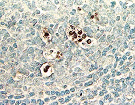
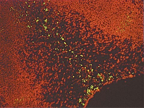
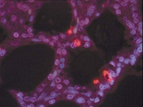
The ApopTag Peroxidase In Situ Apoptosis Detection Kit detects apoptotic cells in situ by labeling & detecting DNA strand breaks by the TUNEL method.
Synonyme(s) :
ApopTag Kit, In Situ Apoptosis Detection Kit
About This Item
Produits recommandés
Niveau de qualité
Fabricant/nom de marque
ApopTag
Chemicon®
Méthode de détection
colorimetric
Conditions d'expédition
dry ice
Description générale
Of all the aspects of apoptosis, the defining characteristic is a complete change in cellular morphology. As observed by electron microscopy, the cell undergoes shrinkage, chromatin margination, membrane blebbing, nuclear condensation and then segmentation, and division into apoptotic bodies which may be phagocytosed (11, 19, 24). The characteristic apoptotic bodies are short-lived and minute, and can resemble other cellular constituents when viewed by brightfield microscopy. DNA fragmentation in apoptotic cells is followed by cell death and removal from the tissue, usually within several hours (7). A rate of tissue regression as rapid as 25% per day can result from apparent apoptosis in only 2-3% of the cells at any one time (6). Thus, the quantitative measurement of an apoptotic index by morphology alone can be difficult.
DNA fragmentation is usually associated with ultrastructural changes in cellular morphology in apoptosis (26, 38). In a number of well-researched model systems, large fragments of 300 kb and 50 kb are first produced by endonucleolytic degradation of higher-order chromatin structural organization. These large DNA fragments are visible on pulsed-field electrophoresis gels (5, 43, 44). In most models, the activation of Ca2+- and Mg2+-dependent endonuclease activity further shortens the fragments by cleaving the DNA at linker sites between nucleosomes (3). The ultimate DNA fragments are multimers of about 180 bp nucleosomal units. These multimers appear as the familiar "DNA ladder" seen on standard agarose electrophoresis gels of DNA extracted from many kinds of apoptotic cells (e.g. 3, 7,13, 35, 44).
Another method for examining apoptosis via DNA fragmentation is by the TUNEL assay, (13) which is the basis of ApopTag technology. The DNA strand breaks are detected by enzymatically labeling the free 3′-OH termini with modified nucleotides. These new DNA ends that are generated upon DNA fragmentation are typically localized in morphologically identifiable nuclei and apoptotic bodies. In contrast, normal or proliferative nuclei, which have relatively insignificant numbers of DNA 3′-OH ends, usually do not stain with the kit. ApopTag Kits detect single-stranded (25) and double-stranded breaks associated with apoptosis. Drug-induced DNA damage is not identified by the TUNEL assay unless it is coupled to the apoptotic response (8). In addition, this technique can detect early-stage apoptosis in systems where chromatin condensation has begun and strand breaks are fewer, even before the nucleus undergoes major morphological changes (4, 8).
Apoptosis is distinct from accidental cell death (necrosis). Numerous morphological and biochemical differences that distinguish apoptotic from necrotic cell death are summarized in the following table (adapted with permission from reference 39).
Composants
Tampon de réaction : 2,0 ml, -15 °C à -25 °C
Enzyme TdT : 0,64 ml, -15 °C à -25 °C
Tampon de blocage/lavage : 20 ml, -15 °C à -25 °C
Anticorps anti-digoxigénine couplés à la peroxydase* : 3,0 ml, 2 °C à 8 °C
Lamelles en plastique : 100 unités, température ambiante
Remarque : DAB (substrat de la peroxydase) non fournie. Elle doit être achetée séparément.
Nombre d'échantillons par kit : Le contenu de ce kit, utilisé conformément aux instructions, permet de colorer 40 échantillons de tissus d'environ 5 mm2 chacun. Si le kit est utilisé pour l'analyse d'échantillons sur lame, le tampon de réaction sera épuisé avant les autres réactifs.
Stockage et stabilité
Précautions
1. Les éléments suivants du kit contiennent du cacodylate de potassium (acide diméthylarsinique) en tant que tampon : tampon d'équilibration (réf. 90416), tampon de réaction (réf. 90417) et enzyme TdT (réf. 90418). Ces éléments sont nocifs en cas d'ingestion ; éviter tout contact avec la peau et les yeux (porter des gants et des lunettes) et rincer immédiatement en cas de contact.
2. Les anticorps conjugués (réf. 90420) et les solutions de blocage (n° 10 et n° 13) contiennent 0,08 % d'azide de sodium en tant qu'agent conservateur.
3. L'enzyme TdT (réf. 90418) contient du glycérol ; elle ne congèle pas à -20 °C. Pour une durée de conservation maximale, ne pas amener ce réactif à température ambiante avant utilisation.
Informations légales
Mention d'avertissement
Danger
Mentions de danger
Conseils de prudence
Classification des risques
Aquatic Chronic 2 - Carc. 1B - Skin Sens. 1 - STOT RE 2 Inhalation
Organes cibles
Respiratory Tract
Code de la classe de stockage
6.1C - Combustible acute toxic Cat.3 / toxic compounds or compounds which causing chronic effects
Certificats d'analyse (COA)
Recherchez un Certificats d'analyse (COA) en saisissant le numéro de lot du produit. Les numéros de lot figurent sur l'étiquette du produit après les mots "Lot" ou "Batch".
Déjà en possession de ce produit ?
Retrouvez la documentation relative aux produits que vous avez récemment achetés dans la Bibliothèque de documents.
Les clients ont également consulté
Articles
Cellular apoptosis assays to detect programmed cell death using Annexin V, Caspase and TUNEL DNA fragmentation assays.
Notre équipe de scientifiques dispose d'une expérience dans tous les secteurs de la recherche, notamment en sciences de la vie, science des matériaux, synthèse chimique, chromatographie, analyse et dans de nombreux autres domaines..
Contacter notre Service technique