Accéder au contenu
MilliporeSigma
Toutes les photos(2)

Key Documents

MABN293

Sigma-Aldrich

Anti-Peripherin-2 Antibody, clone 6B10.1

clone 6B10.1, from mouse

Synonyme(s) :

Peripherin-2, Retinal degeneration slow protein, Tetraspanin-22, Tspan-22

Se connecterpour consulter vos tarifs contractuels et ceux de votre entreprise/organisme


About This Item

Code UNSPSC :
12352203
eCl@ss :
32160702
Nomenclature NACRES :
NA.41

Source biologique

mouse

Niveau de qualité

Forme d'anticorps

purified immunoglobulin

Type de produit anticorps

primary antibodies

Clone

6B10.1, monoclonal

Espèces réactives

mouse, rat, human

Technique(s)

immunohistochemistry: suitable
western blot: suitable

Isotype

IgG1κ

Numéro d'accès NCBI

Numéro d'accès UniProt

Conditions d'expédition

wet ice

Modification post-traductionnelle de la cible

unmodified

Informations sur le gène

human ... PRPH2(5961)

Description générale

Peripherin-2 (PRPH2) is also called Retinal degeneration slow protein (RDS) and Tetraspanin-22 (Tspan-22). Peripherin-2 is a critical factor in ocular disk morphogenesis and possibly functions as an adhesion molecule helping to stabilize and compact outer segment disks, to help maintain curvature of the rim. Peripherin-2 is expressed in the photoreceptors of the retina and in the rim of rod outer segment (ROS) disks. Peripherin-2 defects are found in different retinal diseases including cone-rod dystrophy, retinitis pigmentosa, and macular degeneration.

Immunogène

Epitope: Cytoplasmic domain
GST-tagged recombinant protein corresponding to the cytoplasmic domain of human Peripherin-2.

Application

Anti-Peripherin-2 Antibody, clone 6B10.1 is a highly specific mouse monoclonal antibody, that targets Peripherin & has been tested in western blotting & IHC.
Research Category
Neuroscience
Research Sub Category
Developmental Signaling
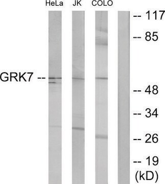
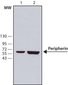
Western Blotting Analysis: A 1:1,000 dilution from a representative lot detected Peripherin-2 in 10 µg of human retina tissue lysate.
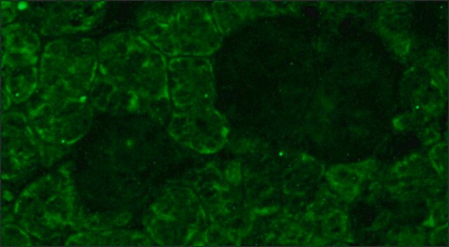
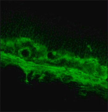
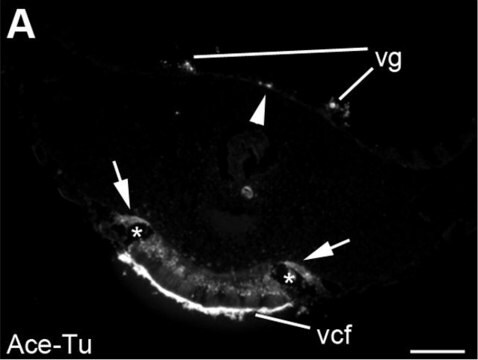
Immunohistochemistry Analysis: A 1:2,000 dilution from a representative lot detected Peripherin-2 in human retina tissue.

Qualité

Evaluated by Western Blotting in mouse eye tissue lysate.

Western Blotting Analysis: A 1:1,000 dilution of this antibody detected Peripherin-2 in 10 µg of mouse eye tissue lysate.

Description de la cible

~35 kDa observed

Forme physique

Format: Purified
Protein G Purified
Purified mouse monoclonal IgG1κ in buffer containing 0.1 M Tris-Glycine (pH 7.4), 150 mM NaCl with 0.05% sodium azide.

Stockage et stabilité

Stable for 1 year at 2-8°C from date of receipt.

Clause de non-responsabilité

Unless otherwise stated in our catalog or other company documentation accompanying the product(s), our products are intended for research use only and are not to be used for any other purpose, which includes but is not limited to, unauthorized commercial uses, in vitro diagnostic uses, ex vivo or in vivo therapeutic uses or any type of consumption or application to humans or animals.

Vous ne trouvez pas le bon produit ?  

Essayez notre Outil de sélection de produits.

Code de la classe de stockage

12 - Non Combustible Liquids

Classe de danger pour l'eau (WGK)

WGK 1

Point d'éclair (°F)

Not applicable

Point d'éclair (°C)

Not applicable


Certificats d'analyse (COA)

Recherchez un Certificats d'analyse (COA) en saisissant le numéro de lot du produit. Les numéros de lot figurent sur l'étiquette du produit après les mots "Lot" ou "Batch".

Déjà en possession de ce produit ?

Retrouvez la documentation relative aux produits que vous avez récemment achetés dans la Bibliothèque de documents.

Consulter la Bibliothèque de documents

Kai Yao et al.
Nature, 560(7719), 484-488 (2018-08-17)
In zebrafish, Müller glia (MG) are a source of retinal stem cells that can replenish damaged retinal neurons and restore vision1. In mammals, however, MG do not spontaneously re-enter the cell cycle to generate a population of stem or progenitor
Katie L Bales et al.
Investigative ophthalmology & visual science, 61(10), 17-17 (2020-08-11)
To identify the role of the BBSome protein Bardet-Biedl syndrome 5 (BBS5) in photoreceptor function, protein trafficking, and structure using a congenital mutant mouse model. Bbs5-/- mice (2 and 9 months old) were used to assess retinal function and morphology.
Suguru Yamasaki et al.
iScience, 25(1), 103657-103657 (2022-01-14)
Pluripotent stem cell (PSC)-derived retinal sheet transplanted in vivo can form structured photoreceptor layers, contact with host bipolar cells, and transmit light signals to host retinas. However, a major concern is the presence of graft bipolar cells that may impede host-graft

Notre équipe de scientifiques dispose d'une expérience dans tous les secteurs de la recherche, notamment en sciences de la vie, science des matériaux, synthèse chimique, chromatographie, analyse et dans de nombreux autres domaines..

Contacter notre Service technique