Skip to Content
MilliporeSigma
All Photos(2)

Key Documents

C1639

Sigma-Aldrich

Collagenase from Clostridium histolyticum

0.2 μm filtered, for general use, Type I-S, 0.2-1.0 FALGPA units/mg solid, ≥125 CDU/mg solid

Synonym(s):

Clostridiopeptidase A

Sign Into View Organizational & Contract Pricing


About This Item

CAS Number:
Enzyme Commission number:
EC Number:
MDL number:
UNSPSC Code:
12352204
NACRES:
NA.54

Quality Level

sterility

0.2 μm filtered

form

lyophilized powder

specific activity

≥125 CDU/mg solid
0.2-1.0 FALGPA units/mg solid

mol wt

68-130 kDa

storage temp.

−20°C

Looking for similar products? Visit Product Comparison Guide

Application

This product is suitable for the disaggregation of human tumor, mouse kidney, human adult and fetal brain, lung and many other epithelia tissues. It has also been shown to be effective in liver and kidney perfusion studies, digestion of pancreas, isolation of nonparenchymal rat liver cells and hepatocyte preparation. Collagenase has also been used in the preparation of arterial tissue for the study of Advanced Glycosylation End Products. This enzyme has been tested for the release of heptatocytes at a concentration of approximately 1 mg/mL. Concentrations for digestion range from 0.1 to 5 mg/mL.

Biochem/physiol Actions

The collagenase product is a mixture of enzymes secreted by C. histolyticum, with different products differentiated by the relative ratios of the 10-18 components found in the secreted enzymes. The main components are two collagenases, clostripain, and a neutral protease. The synergistic action of these enzymes degrade collagen and other intracellular materialThe action of both collagenase enzymes and the neutral protease is necessary for effective release of cells from tissue. Various types of collagen are the natural substrates for collagenase.
Collagenase is activated by four gram atom calcium per mole enzyme. It is inhibited by ethylene glycol-bis(beta-aminoethyl ether) - N, N, N′,N′-tetraacetic acid, beta-mercaptoethanol, glutathione, thioglycolic acid and 8-hydroxyquinoline.

Caution

As supplied, this product is stable for one year at -20°C. There is no loss in FALGPA or protease activity in 30 days at 37°C, 50°C and -20°C. Solutions of crude collagenase are stable if frozen quickly in aliquots (at 10 mg/mL) and kept frozen at -20°C. Further freeze-thaw cycles will damage the solution. The product retains 100% activity over 7 hours when held on ice.

Unit Definition

One collagen digestion unit (CDU) liberates peptides from collagen from bovine achilles tendon equivalent in ninhydrin color to 1.0 μmole of leucine in 5 hours at pH 7.4 at 37 °C in the presence of calcium ions. One FALGPA hydrolysis unit hydrolyzes 1.0 μmole of furylacryloyl-Leu-Gly-Pro-Ala per min at 25°C. One Neutral Protease unit hydrolyzes casein to produce color equivalent to 1.0 μmole of tyrosine per 5 hr at pH 7.5 at 37°C. One Clostripain Unit hydrolyzes 1.0 μmole of BAEE per min at pH 7.6 at 25°C in the presence of DTT.

Preparation Note

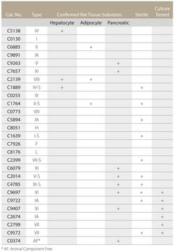
Prepared from Type I (C0130). A stock solution may be prepared by dissolving 0.05-0.1 mg/mL of collagenase in 50 mM TES buffer containing 0.36 mM calcium chloride (TESCA, pH 7.4) at 37 °C.

substrate

Product No.
Description
Pricing

Pictograms

Health hazardExclamation mark

Signal Word

Danger

Hazard Statements

Hazard Classifications

Eye Irrit. 2 - Resp. Sens. 1 - Skin Irrit. 2 - STOT SE 3

Target Organs

Respiratory system

Storage Class Code

11 - Combustible Solids

WGK

WGK 1

Flash Point(F)

Not applicable

Flash Point(C)

Not applicable

Personal Protective Equipment

dust mask type N95 (US), Eyeshields, Gloves

Certificates of Analysis (COA)

Search for Certificates of Analysis (COA) by entering the products Lot/Batch Number. Lot and Batch Numbers can be found on a product’s label following the words ‘Lot’ or ‘Batch’.

Already Own This Product?

Find documentation for the products that you have recently purchased in the Document Library.

Visit the Document Library

Akane Morohoshi et al.
Development (Cambridge, England), 148(21) (2021-10-30)
The acrosome is a cap-shaped, Golgi-derived membranous organelle that is located over the anterior of the sperm nucleus and highly conserved throughout evolution. Although morphological changes during acrosome biogenesis in spermatogenesis have been well described, the molecular mechanism underlying this
Cuiping Zhang et al.
Cell death & disease, 11(6), 470-470 (2020-06-20)
Mesenchymal stem cells (MSCs) have been used in cell-based therapies for a variety of disorders. Some factors such as inflammatory mediators in the diseased area might damage the survival of MSCs and affect their efficacy. Pyroptosis is a form of
Tony Schountz et al.
Viruses, 11(3) (2019-03-27)
Syrian hamsters (Mesocricetus auratus) are a pathogenesis model for the Nipah virus (NiV), and we sought to determine if they are also susceptible to the Cedar virus (CedPV). Following intranasal inoculation with CedPV, virus replication occurred in the lungs and
Hyunji Byun et al.
Reproductive biology and endocrinology : RB&E, 19(1), 112-112 (2021-07-18)
The tumor susceptibility gene 101 (Tsg101), a component of the endosomal sorting complex required for transport (ESCRT) complex I, is involved in multiple biological processes involving endomembranous structures and the plasma membrane. The role of Tsg101 in the uterine epithelium
Ralph Stadhouders et al.
Nature protocols, 8(3), 509-524 (2013-02-16)
Chromosome conformation capture (3C) technology is a powerful and increasingly popular tool for analyzing the spatial organization of genomes. Several 3C variants have been developed (e.g., 4C, 5C, ChIA-PET, Hi-C), allowing large-scale mapping of long-range genomic interactions. Here we describe

Our team of scientists has experience in all areas of research including Life Science, Material Science, Chemical Synthesis, Chromatography, Analytical and many others.

Contact Technical Service