Skip to Content
MilliporeSigma
All Photos(1)

Key Documents

05-716

Sigma-Aldrich

Anti-Myosin Heavy Chain Antibody, clone A4.1025

ascites fluid, clone A4.1025, Upstate®

Synonym(s):

Myosin heavy chain 1, Myosin heavy chain 2x, Myosin heavy chain IIx/d, Myosin heavy chain, skeletal muscle, adult 1, myosin, heavy chain 1, skeletal muscle, adult, myosin, heavy polypeptide 1, skeletal muscle, adult

Sign Into View Organizational & Contract Pricing


About This Item

UNSPSC Code:
12352203
eCl@ss:
32160702
NACRES:
NA.41

biological source

mouse

antibody form

ascites fluid

antibody product type

primary antibodies

clone

A4.1025, monoclonal

species reactivity

mouse, zebrafish, rat, Drosophila, rabbit, human

manufacturer/tradename

Upstate®

technique(s)

immunohistochemistry: suitable
western blot: suitable

isotype

IgG

NCBI accession no.

UniProt accession no.

shipped in

dry ice

target post-translational modification

unmodified

Gene Information

human ... MYH1(4619)

General description

Myosin is one of the two major components of muscle tissue responsible for contraction. In muscle cells myosin is arranged as thick filamentous structures that are arranged in parallel to thin filamentous structures composed of actin. Muscle contraction is actuated by the coupling and uncoupling of thin and thick filaments through a chemical linkage mediated by ATP. The coupling and uncoupling is arranged such that the filaments slide in one direction when coupled and in reverse direction when uncoupled or released. Myosin and actin also serve to facilitate motility in non-muscle cells such as in slime molds where shorter yet similar filamentous structures work together. An increasing number of different myosins are being described. Myosin I is a low molecular weight myosin found in protozoa and does not self-assemble. Myosin II is the classic form found in muscle cells that self assemble into thick bundles. Myosin II is a multimeric protein that is composed of two heavy chains and two light chains.

Specificity

This antibody recognizes myosin heavy chain, Mr ~220 kDa. Immunoblotting with muscle cell lysates will often show several close bands from different myosin heavy chain genes, as well as degradation products.

Immunogen

Partially purified protein from adult human skeletal muscle myosin. Clone A4.1025.

Application

Anti-Myosin Heavy Chain Antibody, clone A4.1025 is an antibody against Myosin Heavy Chain for use in IH & WB.
Immunohistochemistry:
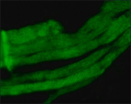
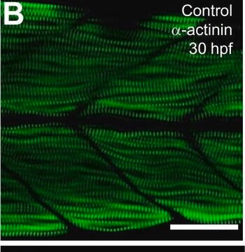
This antibody has been reported by an independent laboratory to detect myosin heavy chain in zebrafish embryos and skeletal muscle sections fixed with 4% paraformaldehyde in a previous lot (Blagden, C.S., 1997; Cho, M., 1994).

Quality

Routinely evaluated by Western Blot on RIPA lysates from C2C12 cells.

Western Blot Analysis:
1:1000-1:5000 dilution of this lot detected Myosin Heavy Chain (MyHC) in RIPA lysates from Rabbit skeletal muscle cells.

Target description

~220 kDa

Physical form

Unpurified mouse ascites monoclonal IgG in buffer containing 0.07 M Tris-glycine, pH 7.4, 0.105 M NaCl, 0.035% sodium azide and 30% glycerol. Liquid at -20°C.

Storage and Stability

Stable for 1 year at -20ºC from date of receipt.
For maximum recovery of product, centrifuge the vial prior to removing the cap.

Handling Recommendations:
Upon first thaw, and prior to removing the cap, centrifuge the vial and gently mix the solution. Aliquot into microcentrifuge tubes and store at -20°C. Avoid repeated freeze/thaw cycles, which may damage IgG and affect product performance. Note: Variability in freezer temperatures below -20°C may cause glycerol-containing solutions to become frozen during storage.

Analysis Note

Control
RIPA lysates from C2C12 cells

Other Notes

Concentration: Please refer to the Certificate of Analysis for the lot-specific concentration.

Legal Information

UPSTATE is a registered trademark of Merck KGaA, Darmstadt, Germany

Not finding the right product?  

Try our Product Selector Tool.

Storage Class Code

10 - Combustible liquids

WGK

WGK 1


Certificates of Analysis (COA)

Search for Certificates of Analysis (COA) by entering the products Lot/Batch Number. Lot and Batch Numbers can be found on a product’s label following the words ‘Lot’ or ‘Batch’.

Already Own This Product?

Find documentation for the products that you have recently purchased in the Document Library.

Visit the Document Library

Effects of doxorubicin cancer therapy on autophagy and the ubiquitin-proteasome system in long-term cultured adult rat cardiomyocytes.
Polychronis Dimitrakis,Maria-Iris Romay-Ogando,Francesco Timolati,Thomas M Suter,Christian Zuppinger
Cell and Tissue Research null
A M Maggs et al.
Journal of muscle research and cell motility, 21(2), 101-113 (2000-08-29)
The contractile properties of muscle fibres are, in part, determined by the myosin heavy chain (MyHC) isoforms they express. Using monoclonal antibodies, we show that at least three forms of slow twitch MyHC accumulate sequentially during mouse fetal development and
Mechanical stress triggers cardiomyocyte autophagy through angiotensin II type 1 receptor-mediated p38MAP kinase independently of angiotensin II.
Lin, L; Tang, C; Xu, J; Ye, Y; Weng, L; Wei, W; Ge, J; Liu, X; Zou, Y
Testing null
Yan-Ping Yang et al.
Development, growth & differentiation, 55(3), 359-367 (2013-03-16)
The second heart field (SHF) is indicated to contribute to the embryonic heart development. However, less knowledge is available about SHF development of human embryo due to the difficulty of collecting embryos. In this study, serial sections of human embryos
Notochord induction of zebrafish slow muscle mediated by Sonic hedgehog
Blagden, C. S., et al
Genes & Development, 11, 2163-2175 (1997)

Our team of scientists has experience in all areas of research including Life Science, Material Science, Chemical Synthesis, Chromatography, Analytical and many others.

Contact Technical Service