Skip to Content
MilliporeSigma
All Photos(5)

Key Documents

HPA001349

Sigma-Aldrich

Anti-MYH6 antibody produced in rabbit

Prestige Antibodies® Powered by Atlas Antibodies, affinity isolated antibody, buffered aqueous glycerol solution

Synonym(s):

Anti-MYH7, Anti-MyHC-α antibody produced in rabbit, Anti-Myosin heavy chain 6 antibody produced in rabbit, Anti-Myosin heavy chain, cardiac muscle α-isoform antibody produced in rabbit, Anti-Myosin-6 antibody produced in rabbit

Sign Into View Organizational & Contract Pricing


About This Item

UNSPSC Code:
12352203
Human Protein Atlas Number:
NACRES:
NA.41

biological source

rabbit

Quality Level

conjugate

unconjugated

antibody form

affinity isolated antibody

antibody product type

primary antibodies

clone

polyclonal

product line

Prestige Antibodies® Powered by Atlas Antibodies

form

buffered aqueous glycerol solution

species reactivity

human

technique(s)

immunohistochemistry: 1:2500-1:5000

immunogen sequence

QVEEDKVNSLSKSKVKLEQQVDDLEGSLEQEKKVRMDLERAKRKLEGDLKLTQESIMDLENDKLQLEEKLKKKEFDINQQNSKIEDEQVLALQLQKKLKENQARIEELEEELEAERTARAKVEKLRSDLSRELEEISERLEEA

UniProt accession no.

shipped in

wet ice

storage temp.

−20°C

target post-translational modification

unmodified

Gene Information

Related Categories

Immunogen

Myosin-6 recombinant protein epitope signature tag (PrEST)

Application

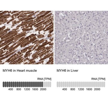
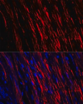
Anti-MYH6 antibody produced in rabbit, a Prestige Antibody, is developed and validated by the Human Protein Atlas (HPA) project . Each antibody is tested by immunohistochemistry against hundreds of normal and disease tissues. These images can be viewed on the Human Protein Atlas (HPA) site by clicking on the Image Gallery link. The antibodies are also tested using immunofluorescence and western blotting. To view these protocols and other useful information about Prestige Antibodies and the HPA, visit sigma.com/prestige.

Biochem/physiol Actions

MYH6 (myosin, heavy chain 6) gene encodes the α heavy chain subunit of the cardiac muscle myosin, which is a hexamer made up of two heavy chain subunits, two light chain subunits, and two regulatory subunits. It is involved in the contraction of muscles. Defects in this gene cause atrial septal defect 3 (ASD3) characterized by incomplete closure of the wall between the atria resulting in blood flow from the left to the right atria. Mutations in this gene also cause familial hypertrophic cardiomyopathy that is characterized by ventricular hypertrophy, dyspnea, syncope, collapse, palpitations, and chest pain.

Features and Benefits

Prestige Antibodies® are highly characterized and extensively validated antibodies with the added benefit of all available characterization data for each target being accessible via the Human Protein Atlas portal linked just below the product name at the top of this page. The uniqueness and low cross-reactivity of the Prestige Antibodies® to other proteins are due to a thorough selection of antigen regions, affinity purification, and stringent selection. Prestige antigen controls are available for every corresponding Prestige Antibody and can be found in the linkage section.

Every Prestige Antibody is tested in the following ways:
  • IHC tissue array of 44 normal human tissues and 20 of the most common cancer type tissues.
  • Protein array of 364 human recombinant protein fragments.

Linkage

Corresponding Antigen APREST70518

Physical form

Solution in phosphate-buffered saline, pH 7.2, containing 40% glycerol and 0.02% sodium azide

Legal Information

Prestige Antibodies is a registered trademark of Merck KGaA, Darmstadt, Germany

Disclaimer

Unless otherwise stated in our catalog or other company documentation accompanying the product(s), our products are intended for research use only and are not to be used for any other purpose, which includes but is not limited to, unauthorized commercial uses, in vitro diagnostic uses, ex vivo or in vivo therapeutic uses or any type of consumption or application to humans or animals.

Not finding the right product?  

Try our Product Selector Tool.

Storage Class Code

10 - Combustible liquids

WGK

WGK 1

Flash Point(F)

Not applicable

Flash Point(C)

Not applicable

Personal Protective Equipment

dust mask type N95 (US), Eyeshields, Gloves

Choose from one of the most recent versions:

Certificates of Analysis (COA)

Lot/Batch Number

Don't see the Right Version?

If you require a particular version, you can look up a specific certificate by the Lot or Batch number.

Already Own This Product?

Find documentation for the products that you have recently purchased in the Document Library.

Visit the Document Library

T A Epp et al.
Genomics, 18(3), 505-509 (1993-12-01)
The human myocardium expresses two cardiac myosin heavy chain (MyHC) isoforms, alpha and beta, that exist in tandem array on chromosome 14q12. We have previously sequenced the entire human cardiac beta-MyHC gene and now report the complete nucleotide sequence of
Yung-Hao Ching et al.
Nature genetics, 37(4), 423-428 (2005-03-01)
Atrial septal defect is one of the most common forms of congenital heart malformation. We identified a new locus linked with atrial septal defect on chromosome 14q12 in a large family with dominantly inherited atrial septal defect. The underlying mutation
Elisa Carniel et al.
Circulation, 112(1), 54-59 (2005-07-07)
Mutations in the beta-myosin heavy-chain (betaMyHC) gene cause hypertrophic (HCM) and dilated (DCM) forms of cardiomyopathy. In failing human hearts, downregulation of alphaMyHC mRNA or protein has been correlated with systolic dysfunction. We hypothesized that mutations in alphaMyHC could also
Morihiko Aoyama et al.
Circulation. Heart failure, 9(1), e002081-e002081 (2016-01-02)
Ample evidence demonstrates cardiovascular protection by incretin-based therapy using dipeptidyl peptidase 4 inhibitor (DPP4i) and glucagon-like peptide-1 (GLP-1) under either diabetic or nondiabetic condition. Their action on myocardium is mediated by the cyclic AMP (cAMP) signal; however, the pathway remains
Nina Tandon et al.
Journal of tissue engineering and regenerative medicine, 5(6), e115-e125 (2011-05-24)
In vitro application of pulsatile electrical stimulation to neonatal rat cardiomyocytes cultured on polymer scaffolds has been shown to improve the functional assembly of cells into contractile engineered cardiac tissues. However, to date, the conditions of electrical stimulation have not

Our team of scientists has experience in all areas of research including Life Science, Material Science, Chemical Synthesis, Chromatography, Analytical and many others.

Contact Technical Service