Z764000
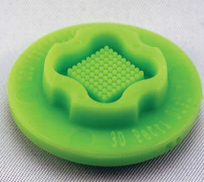
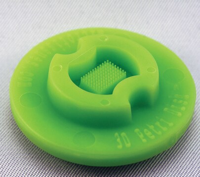
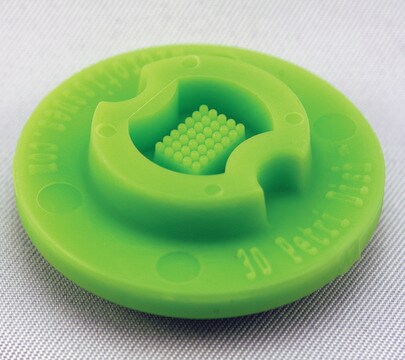
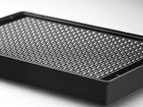
MicroTissues® 3D Petri Dish® micro-mold spheroids
size S, 16 x 16 array, fits 12 well plates
Sinônimo(s):
3D, 3D Cell Culture
Faça loginpara ver os preços organizacionais e de contrato
About This Item
Código UNSPSC:
41121812
NACRES:
NB.14
Produtos recomendados
Materiais
spherical
tamanho
S
esterilidade
sterile; autoclaved
Características
lid: no
16 x 16 array
embalagem
pack of 6 ea
fabricante/nome comercial
MicroTissues Inc. 12-256
volume
190 μL
Procurando produtos similares? Visita Guia de comparação de produtos
Descrição geral
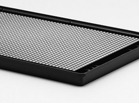
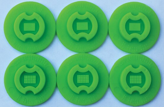
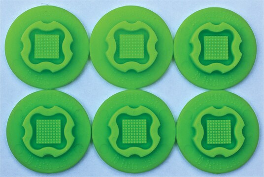
Six autoclavable precision micro-molds to cast 3D Petri Dish for forming small spheroids. 3D Petri Dish for use in 12-well plate. Each micro-mold forms 256 circular recesses in a 16 x 16 array.
- Nominal dimensions of each 3D culture recess: diam. 300 μm x D 800 μm
- Micro-molds also form single chamber for cell seeding
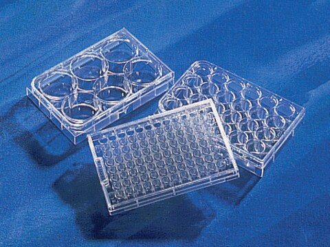
When the gelled agarose is removed from the micro-mold, it is transferred to a standard 12 well or 24 well tissue culture dish and equilibrated with cell culture medium.
Since the agarose is transparent, the spheroids or microtissues that form at the bottom of each agarose micro-well can be easily viewed using a standard inverted microscope using phase contrast, bright field or fluorescence microscopy.
Since the agarose is transparent, the spheroids or microtissues that form at the bottom of each agarose micro-well can be easily viewed using a standard inverted microscope using phase contrast, bright field or fluorescence microscopy.
- The micro-molds are reusable up to 12 times and can be sterilized via a standard steam autoclave (30 min, dry cycle)
- The micro-molds should be stored in a covered container to maintain sterility and to avoid collecting dust or fibres on their small features
- Any microscope with sufficient magnification and resolution functions for cell based views or images will be suitable for use with the 3D Petri Dish products
Informações legais
3D Petri Dish is a registered trademark of MicroTissues Inc.
MicroTissues is a registered trademark of MicroTissues Inc.
Escolha uma das versões mais recentes:
Certificados de análise (COA)
Lot/Batch Number
Lamentamos, não temos COA para este produto disponíveis online no momento.
Se precisar de ajuda, entre em contato Atendimento ao cliente
Já possui este produto?
Encontre a documentação dos produtos que você adquiriu recentemente na biblioteca de documentos.
Os clientes também visualizaram
Minjae Kim et al.
Journal of cell science, 132(19) (2019-09-08)
Cultured rat primitive extraembryonic endoderm (pXEN) cells easily form free-floating multicellular vesicles de novo, exemplifying a poorly studied type of morphogenesis. Here, we reveal the underlying mechanism and the identity of the vesicles. We resolve the morphogenesis into vacuolization, vesiculation
Nossa equipe de cientistas tem experiência em todas as áreas de pesquisa, incluindo Life Sciences, ciência de materiais, síntese química, cromatografia, química analítica e muitas outras.
Entre em contato com a assistência técnica
