Descrição geral
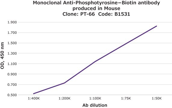
As determined by ELISA and competitive ELISA, the antibody reacts specifically with phosphorylated tyrosine, both as free amino acid or conjugated to carriers such as BSA or KLH. No cross-reactivity is observed with non-phosphorylated tyrosine, phosphothreonine, phosphoserine, AMP or ATP.
Monoclonal Anti-Phosphotyrosine (mouse IgG1 isotype) is derived from the hybridoma produced by the fusion of mouse myeloma cells and splenocytes from an immunized mouse.
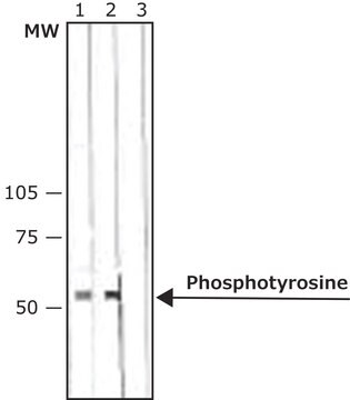
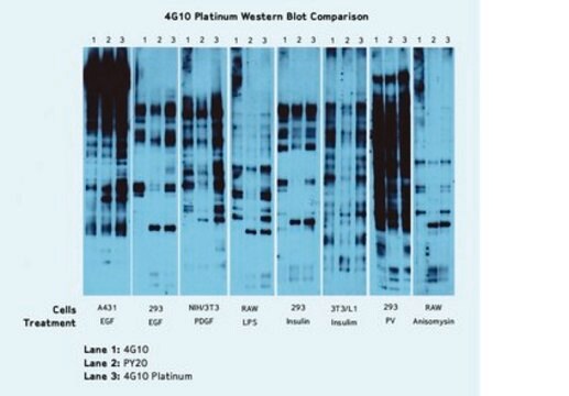
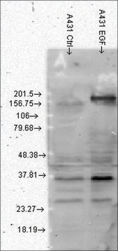
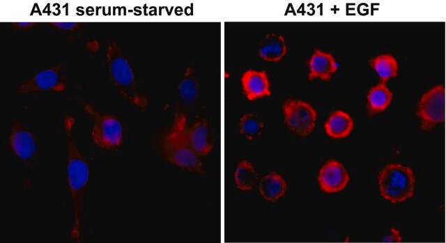
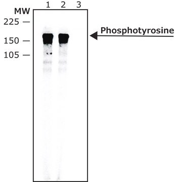
Phosphorylation, attachment of a phosphate group to protein, alters protein functionality by activation or deactivation of the protein, in which protein kinase plays a crucial role. Tyrosine residue phosphorylation plays a key role in cell growth and differentiation. The monoclonal anti-phosphotyrosine antibody is useful in immunoblotting for identification of phosphotyrosine containing protein from cultured human epidermoid carcinoma cell line A-431 and human platelets preparation. This product can be used in immunofluorescent labeling of tyrosine residue at focal adhesion and cellular junctions of cultured MDCK cells. This product is also useful in studies of signal transduction and growth factor receptors. This antibody has shown specificity for binding to proteins that contain phosphorylated tyrosine residue either in free amino acid form or in conjugation with KLH or BSA but will not react with non-phosphorylated tyrosine or other phosphorylated proteins and amino acids. It will also not react with phosphorylated molecules like ATP or AMP. Monoclonal anti-phosphotyrosine reacts specifically with mouse and human.
Especificidade
This antibody is specific for phosphorylated tyrosine both as the free amino acid or when conjugated to carriers such as BSA or KLH
Imunogênio
phosphotyrosine conjugated to BSA
Aplicação
Monoclonal Anti-Phosphotyrosine antibody produced in mouse has been used in
- immunocytochemistry
- immunoprecipitation
- western blotting
- peptide binding studies flow cytometry
- enzyme linked immunosorbent assay (ELISA)
- radioimmunoassay (RIA)
- immunoaffinity isolation
Monoclonal anti-phosphotyrosine antibody produced in mouse is used in immunohistochemisty, flow cytometry, immunoprecipitation, immunoblotting, ELISA and RIA for localization of phosphorylated tyrosine containing proteins. It can also be used for immunoaffinity isolation.
Ações bioquímicas/fisiológicas
Phosphotyrosine levels enhanced in cellular proteins leads to activation of several cellular processes mediated by phosphotyrosine kinases. Phosphotyrosine residues that are autophosphorylated by the binding of ligands associated with receptors like epidermal growth factor (EGF), platelet-derived growth factor (PDGF) and insulin receptors, functions as the primary integral component of mitogenic signaling cascade. Phosphotyrosine mediates the activation of T-cell as a result of phosphorylation of tyrosine residues by tyrosine kinases in cytoplasmic domains of CD4 an CD8 which in turn phosphorylates TCR-CD3 complex. Phosphotyrosine activity has also been found in many retroviruses whose oncogenes encodes tyrosine-specific protein kinase.
Tyrosine residue phosphorylation plays a key role in cell growth and differentiation.
forma física
This product is supplied as ascites fluid containing 15 mM sodium azide
Armazenamento e estabilidade
For continuous use, store at 2-8 °C for up to one month. For extended storage, the solution may be frozen in working aliquots. Repeated freezing and thawing is not recommended. Storage in "frost-free" freezers is not recommended. If slight turbidity occurs upon prolonged storage, clarify the solution by centrifugation before use.
Exoneração de responsabilidade
Unless otherwise stated in our catalog or other company documentation accompanying the product(s), our products are intended for research use only and are not to be used for any other purpose, which includes but is not limited to, unauthorized commercial uses, in vitro diagnostic uses, ex vivo or in vivo therapeutic uses or any type of consumption or application to humans or animals.