Pular para o conteúdo
Merck
Todas as fotos(5)

Documentos Principais

MABT831

Sigma-Aldrich

Anti-Lamin B Receptor (LBR) Antibody, clone BB2SS3F3

clone BB2SS3F3, from mouse

Sinônimo(s):

LMN2R, Integral nuclear envelope inner membrane protein, LBR

Faça loginpara ver os preços organizacionais e de contrato


About This Item

Código UNSPSC:
12352203
eCl@ss:
32160702
NACRES:
NA.41

fonte biológica

mouse

Nível de qualidade

forma do anticorpo

purified immunoglobulin

tipo de produto de anticorpo

primary antibodies

clone

BB2SS3F3, monoclonal

reatividade de espécies

mouse, human

embalagem

antibody small pack of 25 μg

técnica(s)

immunocytochemistry: suitable
immunofluorescence: suitable
western blot: suitable

Isotipo

IgG1κ

nº de adesão NCBI

nº de adesão UniProt

Condições de expedição

ambient

modificação pós-traducional do alvo

unmodified

Informações sobre genes

human ... LBR(3930)

Descrição geral

Lamin-B receptor (UniProt: Q14739; also known as Integral nuclear envelope inner membrane protein, LMN2R) is encoded by the LBR gene (Gene ID: 3930) in human. Lamin-B receptor is a multi-pass membrane protein that anchors the lamina and the heterochromatin to the inner nuclear membrane. It is an integral membrane protein of the interphase nuclear envelope (NE). It contains a Tudor damain that lies within the first 62 amino acids. Its N-terminal end resides in the nucleoplasm, binding to lamin B and heterochromatin and the C-terminal end resides within the inner nuclear membrane, retreating with the endoplasmic reticulum (ER) away from condensing chromosomes during mitotic NE breakdown. During mitosis, Lamin-B receptor is retained within the retreating ER and kept out of the way of the mitotic apparatus. Lamin- B receptor is suggested to perform three vital functions in the cell: (a) contributes to post-mitotic nuclear reformation; (b) contributes to interphase NE growth; (c) contributes to compartmentalization of heterochromatin within the interphase nucleus. Lamin-B receptor is phosphorylated by Cdk1 in mitosis when the inner nuclear membrane breaks down into vesicles that dissociate from the lamina and the chromatin. It is phosphorylated by different protein kinases in interphase when the membrane is associated with these structures. Mutations in LBR gene are known to cause Pelger-Huet anomaly that is characterized by skeletal anomalies, developmental delay, and seizures. Some mutations cause Greenberg dysplasia that can lead to in utero lethality. (Ref.: Olins, AL., et al. (2010). Nucleus 1(1); 53-70).

Especificidade

Clone BB2SS3F3 detects Lamin B receptor in human cells. It targets an epitope within the first 211 amino acids.

Imunogênio

GST-tagged recombinant fragment corresponding to the first 211 amino acids from human Lamin B receptor.

Aplicação

Anti-Lamin B Receptor (LBR), clone BB2SS3F3, Cat. No. MABT831, is a mouse monoclonal antibody that detects Lamin B receptor and has been tested for use in Immunocytochemistry, Immunofluorescence, and Western Blotting.
Immunofluorescence Analysis: A representative lot detected Lamin B Receptor (LBR) in C57BL/6 mouse small intestine (Courtesy of Dr. Brian Burke, Institute of Medical Biology, A*STAR, Singapore).

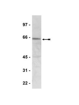
Western Blotting Analysis: A representative lot detected Lamin B Receptor (LBR) in HeLa cell lysate, mouse C2C12 cell lysate, and in adult mouse fibroblasts (MAFs) either LBR+/+ or LBR -/- (Courtesy of Dr. Brian Burke, Institute of Medical Biology, A*STAR, Singapore).

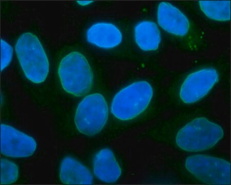
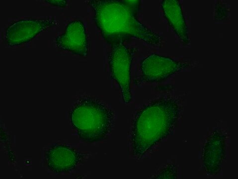
Immunocytochemistry Analysis: A 1:50 dilution from a representative lot detected Lamin B Receptor (LBR) in HeLa cells.

Immunocytochemistry Analysis: A representative lot detected Lamin B Receptor (LBR) in HeLa and NIE-115 cells, as well as in mouse adult fibroblasts (MAF LBR+/+ vs MAF LBR-/-) (Courtesy of Dr. Brian Burke, Institute of Medical Biology, A*STAR, Singapore).
Research Category
Cell Structure

Qualidade

Evaluated by Western Blotting in mouse spleen tissue lysate.

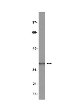
Western Blotting Analysis: 2 µg/mL of this antibody detected Lamin B Receptor (LBR) in 10 µg of mouse spleen tissue lysate.

Descrição-alvo

~55 kDa observed; 70.73 kDa calculated. Uncharacterized bands may be observed in some lysate(s).

forma física

Format: Purified
Protein G purified
Purified mouse monoclonal antibody IgG1 in buffer containing 0.1 M Tris-Glycine (pH 7.4), 150 mM NaCl with 0.05% sodium azide.

Armazenamento e estabilidade

Stable for 1 year at 2-8°C from date of receipt.

Outras notas

Concentration: Please refer to lot specific datasheet.

Exoneração de responsabilidade

Unless otherwise stated in our catalog or other company documentation accompanying the product(s), our products are intended for research use only and are not to be used for any other purpose, which includes but is not limited to, unauthorized commercial uses, in vitro diagnostic uses, ex vivo or in vivo therapeutic uses or any type of consumption or application to humans or animals.

Não está encontrando o produto certo?  

Experimente o nosso Ferramenta de seleção de produtos.

Código de classe de armazenamento

12 - Non Combustible Liquids

Classe de risco de água (WGK)

WGK 1

Ponto de fulgor (°F)

does not flash

Ponto de fulgor (°C)

does not flash


Certificados de análise (COA)

Busque Certificados de análise (COA) digitando o Número do Lote do produto. Os números de lote e remessa podem ser encontrados no rótulo de um produto após a palavra “Lot” ou “Batch”.

Já possui este produto?

Encontre a documentação dos produtos que você adquiriu recentemente na biblioteca de documentos.

Visite a Biblioteca de Documentos

Nossa equipe de cientistas tem experiência em todas as áreas de pesquisa, incluindo Life Sciences, ciência de materiais, síntese química, cromatografia, química analítica e muitas outras.

Entre em contato com a assistência técnica