Pular para o conteúdo
Merck
Todas as fotos(1)

Documentos Principais

MABT340

Sigma-Aldrich

Anti-Proteinase 3/PR3 Antibody, clone MCPR3-2

clone MCPR3-2, from mouse

Sinônimo(s):

Myeloblastin, AGP7, C-ANCA antigen, Leukocyte proteinase 3, Neutrophil proteinase 4, NP-4, P29, PR-3, PR3, Wegener autoantigen

Faça loginpara ver os preços organizacionais e de contrato


About This Item

Código UNSPSC:
12352203
eCl@ss:
32160702
NACRES:
NA.41

fonte biológica

mouse

Nível de qualidade

forma do anticorpo

purified antibody

tipo de produto de anticorpo

primary antibodies

clone

MCPR3-2, monoclonal

reatividade de espécies

human

não deve reagir com

mouse

técnica(s)

ELISA: suitable
affinity binding assay: suitable
flow cytometry: suitable
western blot: suitable

Isotipo

IgG1κ

nº de adesão NCBI

nº de adesão UniProt

Condições de expedição

wet ice

modificação pós-traducional do alvo

unmodified

Informações sobre genes

human ... PRTN3(5657)

Descrição geral

Myeloblastin (EC 3.4.21.76; UniProt P24158; also known as AGP7, C-ANCA antigen, Leukocyte proteinase 3, Neutrophil proteinase 4, NP-4, P29, PR-3, PR3, Wegener autoantigen) is encoded by the PRTN3 (also known as CANCA, MBN, NP4) gene (Gene ID 5657) in human. Proteinase 3 (PR3) is one of four neutral serine proteases (elastase, cathepsin G, PR3, and neutrophil serine protease 4) stored as fully processed mature enzymes in azurophil granules of human neutrophils. Instead of being targeted to granules after their synthesis, some PR3 molecules end up on the surface of neutrophil plasma membrane in either pro- or mature form. The degree of such surface expression is genetically determined, but the surface exposure and pericellular activity of PR3 around neutrophils can be further upregulated upon neutrophil priming and activation. PR3 autoantibody (anti-neutrophil cytoplasmic antibodies or ANCA) is the main pathogenic feature in patients suffering from granulomatosis with polyangiitis (GPA; formerly called Wegener granulomatosis). ANCAs are shown to activate cytokine-primed neutrophils in vitro by cross-linking surface-exposed PR3 and Fcγ receptors. PR3 activity and/or its inactivation by alpha 1-antitrypsin/alpha-1 proteinase inhibitor (α1PI) varies in the human population and contributes to the risk for GPA manifestations either at onset, during relapses, or during systemic progression. PR3 is initially produced with a signal peptide sequence (a.a. 1-25), an N-terminal and a C-terminal propeptide sequence (a.a. 26-27 and 249-256, respectively), the removal of which yields the mature enzyme (a.a. 28-248).

Especificidade

Clone MCPR3-2 detected wild-type PR3 and the catalytically inactive S176A PR3 mutant, but not neutrophil elastase or cathepsin G. Clone MCPR3-2 did not interfere with PR3 enzymatic activity or its interaction with α1-protease inhibitor/α1-PI (Sun, J., et al. (1998). J. Immunol. Methods. 211(1-2):111-123). The ratios of OD readings obtained with PR3-complexed clone MCPR3-2 to the corresponding ODs obtained with PR3-complexed clone MCPR3-3 (Cat. No. MABF973) in ELISA-based serum PR3 autoantibodies (ANCAs) measurements correlate well with the neutralizing activity of ANCAs in patients serum samples (Silva, F., et al. (2010). J. Autoimmun. 35(4) 299-308).

Imunogênio

Epitope: “North-East” of the substrate-binding pocket and adjacent to clone MCPR3-3 epitope (Silva, F., et al. (2010). J. Autoimmun. 35(4) 299-308).
Granule extract from HMC-1/PR3 mast cells expressing transfected human neutrophil PR3 (Sun, J., et al. (1998). J. Immunol. Methods. 211(1-2):111-123).

Aplicação

Anti-Proteinase 3/PR3 Antibody, clone MCPR3-2 is an antibody against PR3 for use in Affinity Binding Assay, ELISA, Flow Cytometry, and Western Blotting.
Research Category
Cell Structure
Research Sub Category
Inflammation & Autoimmune Mechanisms
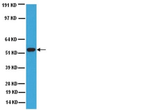
Western Blotting Analysis: 2 µg/mL from a representative lot detected 5 µg of purified human neutrophil PR3 under both non-reducing and reducing conditions (Courtesy of Amber Hummel, Mayo Clinic, Rochester, MN).
Flow Cytometry Analysis: A representative lot detected a large population of human peripheral blood neutrophils with surface PR3. The entire population became positively stained after TNFa-stimulation (Hinkofer, L.C., et al. (2013). J. Biol. Chem. 288(37):26635-26648).
Flow Cytometry Analysis: A representative lot bound immobilized recombinant human PR3 via a distinct epitope than those recognized by clone MCPR3-3 and MCPR3-7 (Cat. No. MABF973 and MABT403, respectively) as determined by FACS analysis of bead-based competition binding assay (Silva, F., et al. (2010). J. Autoimmun. 35(4) 299-308).
Flow Cytometry Analysis: A representative lot bound recombinant human PR3-, but not murine PR3-, coated Talon-beads as determined by FACS analysis. (Silva, F., et al. (2010). J. Autoimmun. 35(4) 299-308).
ELISA Analysis: Representative lots were immobilized on well surface and employed to capture purified human polymorphonuclear cell PR3 or recombinant human PR3 S176A mutant, followed by affinity pull-down of PR3 autoantibodies (anti-neutrophil cytoplasmic antibodies or ANCA) from patients serum samples by the captured PR3 and the subsequent detection of the bound ANCA by alkaline phosphatase-conjugated goat anti-human IgG (Silva, F., et al. (2010). J. Autoimmun. 35(4) 299-308; Sun, J., et al. (1998). J. Immunol. Methods. 211(1-2):111-123).
Western Blotting Analysis: A representative lot detected an exogenously expressed S176A mutant PR3 in lysates from transfected HMC-1 human mast cells as well as purified PR3 from human polymorphonuclear cells (Sun, J., et al. (1998). J. Immunol. Methods. 211(1-2):111-123).
Affinity Binding Assay: A representative lot captured a recombinant human PR3 construct proP-PR3ctp that adopts a pro-PR3 conformation and a recombinant construct ΔPR3ctp-S195A that adopts a mature PR3 conformation with similar affinity (Hinkofer, L.C., et al. (2013). J. Biol. Chem. 288(37):26635-26648).

Qualidade

Identity Confirmation by Isotyping Test.

Isotyping Analysis: The identity of this monoclonal antibody is confirmed by isotyping test to be IgG1κ.

Descrição-alvo

~29 kDa observed. 27.81 kDa (Pro-PR3; a.a. 1-256), 25.14 kDa (N-terminal signal and propeptide removed; a.a. 28-256), 24.25 kDa (Mature; a.a. 28-248) calculated.

forma física

Format: Purified
Protein G Purified
Purified mouse monoclonal IgG1κ antibody in buffer containing 0.1 M Tris-Glycine (pH 7.4), 150 mM NaCl with 0.05% sodium azide.

Armazenamento e estabilidade

Stable for 1 year at 2-8°C from date of receipt.

Outras notas

Concentration: Please refer to lot specific datasheet.

Exoneração de responsabilidade

Unless otherwise stated in our catalog or other company documentation accompanying the product(s), our products are intended for research use only and are not to be used for any other purpose, which includes but is not limited to, unauthorized commercial uses, in vitro diagnostic uses, ex vivo or in vivo therapeutic uses or any type of consumption or application to humans or animals.

Não está encontrando o produto certo?  

Experimente o nosso Ferramenta de seleção de produtos.

Código de classe de armazenamento

12 - Non Combustible Liquids

Classe de risco de água (WGK)

WGK 1

Ponto de fulgor (°F)

Not applicable

Ponto de fulgor (°C)

Not applicable


Certificados de análise (COA)

Busque Certificados de análise (COA) digitando o Número do Lote do produto. Os números de lote e remessa podem ser encontrados no rótulo de um produto após a palavra “Lot” ou “Batch”.

Já possui este produto?

Encontre a documentação dos produtos que você adquiriu recentemente na biblioteca de documentos.

Visite a Biblioteca de Documentos

Nossa equipe de cientistas tem experiência em todas as áreas de pesquisa, incluindo Life Sciences, ciência de materiais, síntese química, cromatografia, química analítica e muitas outras.

Entre em contato com a assistência técnica