Accéder au contenu
Merck
Toutes les photos(1)

Principaux documents

SAB4200742

Sigma-Aldrich

Anti-c-Myc-Peroxidase antibody, Mouse monoclonal

clone 9E10, purified from hybridoma cell culture

Synonyme(s) :

Anti-MRTL, Anti-Myc, Anti-bHLHe39, Anti-v-myc avian myelocytomatosis viral oncogene homolog

Se connecterpour consulter vos tarifs contractuels et ceux de votre entreprise/organisme


About This Item

Code UNSPSC :
12352203
Nomenclature NACRES :
NA.56

Source biologique

mouse

Niveau de qualité

Conjugué

peroxidase conjugate

Forme d'anticorps

purified from hybridoma cell culture

Type de produit anticorps

primary antibodies

Clone

9E10, monoclonal

Forme

lyophilized powder

Espèces réactives

human

Conditionnement

vial of 100 μL

Concentration

~2 mg/mL

Technique(s)

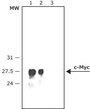
immunoblotting: 1:250-1:500 using lysate of HEK-293T cells over expressing c-Myc fusion protein
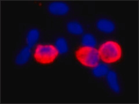
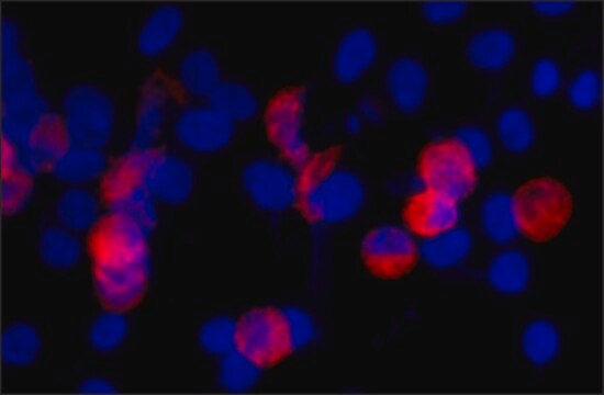
immunohistochemistry: suitable

Isotype

IgG1

Numéro d'accès UniProt

Conditions d'expédition

dry ice

Température de stockage

−20°C

Modification post-traductionnelle de la cible

unmodified

Informations sur le gène

human ... MYC(4609)

Description générale

Monoclonal Anti-c-Myc (mouse IgG1 isotype) is derived from the 9E10 hybridoma, produced by the fusion of mouse myeloma cells and splenocytes from a BALB/c mouse immunized with synthetic peptide of the human p62c-myc protein, conjugated to KLH. c-Myc is a proto-oncogene localized on long arm of human chromosome 8. The encoded protein contains an N-terminal transactivation domain. The human c-Myc proto-oncogene is the cellular homolog of the avian v-Myc gene found in several leukemogenic retroviruses.

Spécificité

Monoclonal Anti-Calponin specifically recognizes smooth muscle Calponin.

Immunogène

Synthetic peptide of the human p62c-myc protein.

Application

Anti-c-Myc-Peroxidase antibody, Mouse monoclonal may be used in immunoblotting and immunohistology.

Actions biochimiques/physiologiques

c-Myc is a transcription factor involved in regulation of cell growth, proliferation, differentiation and apoptosis. It is implicated in DNA replication, RNA splicing or transcription. Increased expression of the gene is associated with the development of various types of human cancers.

Forme physique

Supplied as lyophilized powder. After reconstitution with 0.1 mL of distilled water to a final antibody concentration of ∼ 2 mg/mL, the solution contains 1% BSA, 2.5% trehalose, 0.05% MIT in 0.01 M sodium phosphate buffered saline

Stockage et stabilité

Store the lyophilized product at 2–8 °C. For extended storage after reconstitution, keep at –20 °C in working aliquots. Avoid repeated freeze-thaw cycles. For continuous use after reconstitution, keep at 2–8 °C for up to 1 month. Solutions at working dilution should be discarded if not used within 12 hours.

Clause de non-responsabilité

Unless otherwise stated in our catalog, our products are intended for research use only and are not to be used for any other purpose, which includes but is not limited to, unauthorized commercial uses, in vitro diagnostic uses, ex vivo or in vivo therapeutic uses or any type of consumption or application to humans or animals.

Vous ne trouvez pas le bon produit ?  

Essayez notre Outil de sélection de produits.

Pictogrammes

Exclamation mark

Mention d'avertissement

Warning

Mentions de danger

Classification des risques

Skin Sens. 1

Code de la classe de stockage

13 - Non Combustible Solids

Classe de danger pour l'eau (WGK)

WGK 2

Point d'éclair (°F)

Not applicable

Point d'éclair (°C)

Not applicable


Faites votre choix parmi les versions les plus récentes :

Certificats d'analyse (COA)

Lot/Batch Number

Vous ne trouvez pas la bonne version ?

Si vous avez besoin d'une version particulière, vous pouvez rechercher un certificat spécifique par le numéro de lot.

Déjà en possession de ce produit ?

Retrouvez la documentation relative aux produits que vous avez récemment achetés dans la Bibliothèque de documents.

Consulter la Bibliothèque de documents

K Alitalo et al.
Nature, 306(5940), 274-277 (1983-11-17)
The myelocytomatosis viruses are a family of replication-defective avian retroviruses that cause a variety of tumours in chickens and transform both fibroblasts and macrophages in culture through the activity of their oncogene v-myc. A closely related gene (c-myc) is found
S Pelengaris et al.
Current opinion in genetics & development, 10(1), 100-105 (2000-02-19)
The protein products of many dominant oncogenes are capable of inducing both cell proliferation and apoptosis. Recent experiments employing transgenic mice that express an ectopically regulatable myc gene or protein have begun to elucidate the role of the balance between
Q Hu et al.
Science (New York, N.Y.), 268(5207), 100-102 (1995-04-07)
Phosphatidylinositol (Pl)-3 kinase is one of many enzymes stimulated by growth factors. A constitutively activated mutant, p110, that functions independently of growth factor stimulation was constructed to determine the specific responses regulated by Pl-3 kinase. The p110 protein exhibited high
Ann-Marie Baker et al.
Histopathology, 69(2), 222-229 (2016-01-31)
Recent attempts to study MYC distribution in human samples have been confounded by a lack of agreement in immunohistochemical staining between antibodies targeting the N-terminus and those targeting the C-terminus of the MYC protein. The aim of this study was
D Robertson et al.
The journal of histochemistry and cytochemistry : official journal of the Histochemistry Society, 43(5), 471-480 (1995-05-01)
To determine the ultrastructural distribution of H-ras, the rho proteins rho-A, rho-B, rho-C, and the rac1 protein (members of the ras GTP-binding protein family), we used cDNA expression plasmids in which a short sequence coding for the epitope recognized by

Notre équipe de scientifiques dispose d'une expérience dans tous les secteurs de la recherche, notamment en sciences de la vie, science des matériaux, synthèse chimique, chromatographie, analyse et dans de nombreux autres domaines..

Contacter notre Service technique