Accéder au contenu
Merck
Toutes les photos(1)

Principaux documents

C6930

Sigma-Aldrich

Monoclonal Anti-Cytokeratin Peptide 19 antibody produced in mouse

clone A53-B/A2, tissue culture supernatant

Synonyme(s) :

Anti-CK19, Anti-K19, Anti-K1CS

Se connecterpour consulter vos tarifs contractuels et ceux de votre entreprise/organisme


About This Item

Numéro MDL:
Code UNSPSC :
12352203
Nomenclature NACRES :
NA.41
Clone:
A53-B/A2, monoclonal
application:
ARR
IF
Espèces réactives:
human
Technique(s):
indirect immunofluorescence: 1:50 using formalin-fixed, paraffin-embedded, human tissue sections
microarray: suitable
citations:
13

Source biologique

mouse

Conjugué

unconjugated

Forme d'anticorps

tissue culture supernatant

Type de produit anticorps

primary antibodies

Clone

A53-B/A2, monoclonal

Poids mol.

antigen 40 kDa

Contient

15 mM sodium azide

Espèces réactives

human

Technique(s)

indirect immunofluorescence: 1:50 using formalin-fixed, paraffin-embedded, human tissue sections
microarray: suitable

Isotype

IgG2a

Numéro d'accès UniProt

Conditions d'expédition

dry ice

Température de stockage

−20°C

Modification post-traductionnelle de la cible

unmodified

Informations sur le gène

human ... KRT19(3880)

Description générale

Monoclonal Anti-Cytokeratin Peptide 19 (mouse IgG2a isotype) is derived from the A53-B/A2 hybridoma produced by the fusion of mouse myeloma cells and splenocytes from BALB/c mice immunized with mammary human carcinoma cell line MCF-7. Monoclonal Anti-Cytokeratin Peptide 19, known as clone A53-B/A2, or clone No. Ks 19.1, reacts with the rod domain of human cytokeratin peptide 19 (40 kDa), a cytoskeletal protein restricted to epithelial and carcinoma cells. Cytokeratin 19 is a member of the type I acidic subfamily. It is differentially expressed in various human tissues.

Spécificité

Also cited as clone no. KS 19.1, the antibody reacts with the rod domain of cytokeratin peptide 19, a cytoskeletal protein restricted to epithelial and carcinoma cells. Species cross-reactivity is low or absent. The antibody labels simple epithelia and basal cells of non-cornifying stratified squamous epithelia. It is a useful tool in discriminating simple carcinomas from those of different origin and for carcinoma subtyping. This antibody has also been shown to be a marker of pre-malignant lesions of the oral epithelium.

Immunogène

human mammary carcinoma cell line MCF-7.

Application

Monoclonal Anti-Cytokeratin Peptide 19 antibody produced in mouse is also suitable:
  • for immunoblotting, for immunocytochemistry,
  • for immunofluorescence,
  • as a marker of premalignant lesions of the oral epithelium, to label cytokeratin in formalin-fixed or Carnoy-fixed, paraffin embedded tissue and in frozen sections of human tissue, to label simple epithelia and basal cells of noncornifying stratified squamous epithelia.

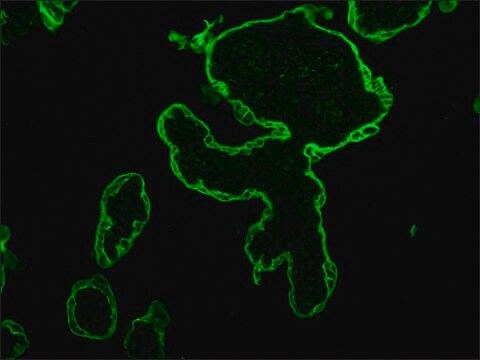
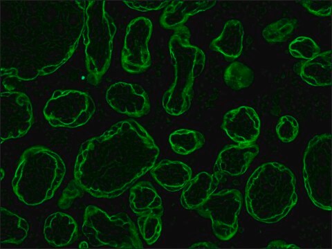
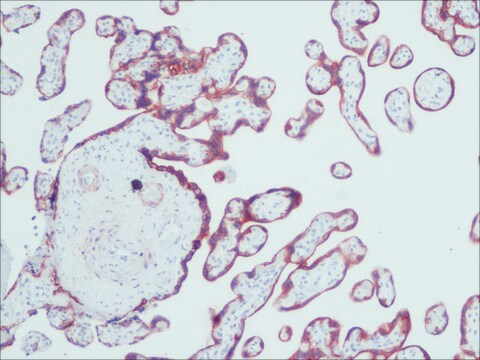
Monoclonal Anti-Cytokeratin Peptide 19 antibody produced in mouse is suitable for microarray and indirect immunofluorescence at a dilution of 1:50 using formalin-fixed, paraffin-embedded and human tissue sections.

Actions biochimiques/physiologiques

Keratin, type I cytoskeletal 19 (40kDa protein) is also known as cytokeratin-19 (CK-19) or keratin-19 (K19). It is encoded by the KRT19 gene in humans. Keratin 19 is a type I keratin. Keratins are intermediate filament-forming proteins that provide mechanical support and perform a variety of additional functions in epithelial cells. K19 is expressed in a subset of hepatocellular carcinomas (HCC) with poor prognosis and is considered as a biliary/hepatic progenitor cell (HPC) marker. It is also a predictor of poorer prognosis for patients with human lung squamous cell carcinoma (SCC).
Monoclonal anti-cytokeratins are specific markers of epithelial cell differentiation and have been widely used as tools in tumor identification and classification.

Clause de non-responsabilité

Unless otherwise stated in our catalog or other company documentation accompanying the product(s), our products are intended for research use only and are not to be used for any other purpose, which includes but is not limited to, unauthorized commercial uses, in vitro diagnostic uses, ex vivo or in vivo therapeutic uses or any type of consumption or application to humans or animals.

Vous ne trouvez pas le bon produit ?  

Essayez notre Outil de sélection de produits.

Code de la classe de stockage

10 - Combustible liquids

Classe de danger pour l'eau (WGK)

nwg

Point d'éclair (°F)

Not applicable

Point d'éclair (°C)

Not applicable


Faites votre choix parmi les versions les plus récentes :

Certificats d'analyse (COA)

Lot/Batch Number

Vous ne trouvez pas la bonne version ?

Si vous avez besoin d'une version particulière, vous pouvez rechercher un certificat spécifique par le numéro de lot.

Déjà en possession de ce produit ?

Retrouvez la documentation relative aux produits que vous avez récemment achetés dans la Bibliothèque de documents.

Consulter la Bibliothèque de documents

Kasper, M., et al.
European Journal of Cancer, 23, 137-137 (1987)
Izabela Podgorski et al.
The American journal of pathology, 175(3), 1255-1269 (2009-08-25)
Bone metastasis is a hallmark of advanced prostate and breast cancers, yet the critical factors behind attraction of tumors to the skeleton have not been validated. Here, we investigated the involvement of cathepsin K in the progression of prostate tumors
Minati Satpathy et al.
Theranostics, 9(3), 778-795 (2019-02-28)
Cancer heterogeneity and drug resistance limit the efficacy of cancer therapy. To address this issue, we have developed an integrated treatment protocol for effective treatment of heterogeneous ovarian cancer. Methods: An amphiphilic polymer coated magnetic iron oxide nanoparticle was conjugated
Targeted drug delivery and image-guided therapy of heterogeneous ovarian cancer using HER2-targeted theranostic nanoparticles
Satpathy M, et al.
Theranostics, 9(3), 778-778 (2019)
R Moll et al.
Laboratory investigation; a journal of technical methods and pathology, 65(1), 74-86 (1991-07-01)
The expression of intermediate filament proteins, particularly individual cytokeratins (CKs), vimentin, and glial filament protein, was immunohistochemically investigated using frozen sections and Carnoy-fixed, paraffin-embedded tissue from normal fetal and adult human kidneys as well as from pathologically altered kidneys. In

Notre équipe de scientifiques dispose d'une expérience dans tous les secteurs de la recherche, notamment en sciences de la vie, science des matériaux, synthèse chimique, chromatographie, analyse et dans de nombreux autres domaines..

Contacter notre Service technique