Accéder au contenu
Merck
Toutes les photos(3)

Key Documents

C0715

Sigma-Aldrich

Monoclonal Anti-Cathepsin D antibody produced in mouse

clone CTD-19, ascites fluid

Synonyme(s) :

Anti-CLN10, Anti-CPSD, Anti-HEL-S-130P

Se connecterpour consulter vos tarifs contractuels et ceux de votre entreprise/organisme


About This Item

Numéro MDL:
Code UNSPSC :
12352203
Nomenclature NACRES :
NA.41

Source biologique

mouse

Niveau de qualité

Conjugué

unconjugated

Forme d'anticorps

ascites fluid

Type de produit anticorps

primary antibodies

Clone

CTD-19, monoclonal

Poids mol.

antigen 34 kDa
antigen 52 kDa (weaker band)

Espèces réactives

human

Technique(s)

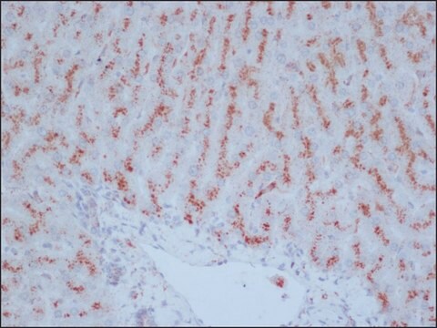
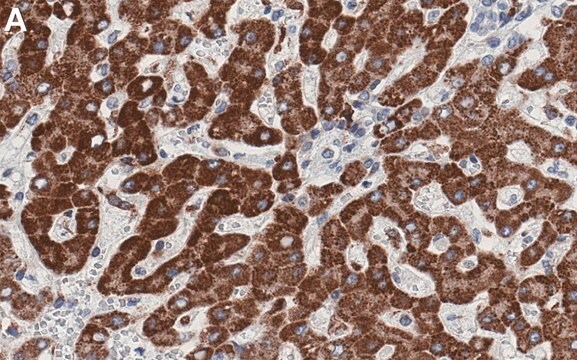
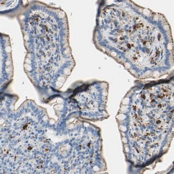
immunohistochemistry (formalin-fixed, paraffin-embedded sections): 1:200 using human breast carcinoma tissue
indirect ELISA: suitable
microarray: suitable
western blot: 1:1,000 using human breast carcinoma cell line extract

Isotype

IgG2a

Numéro d'accès UniProt

Conditions d'expédition

dry ice

Température de stockage

−20°C

Modification post-traductionnelle de la cible

unmodified

Informations sur le gène

human ... CTSD(1509)

Description générale

Cathepsins are lysosomal proteases that play an important role in the intracellular degradation of exogenous and endogenous proteins, in activation of enzyme precursors, and in tumor invasion and metastasis. They are normally localized in lysosomes of almost all mammalian cells, but under certain conditions they can be secreted from the cells.
Cathepsin D (CD, EC 3.4.23.5), an aspartyl endopeptidase, is induced by estrogen in certain estrogen receptor (ER)-positive breast cancer cell lines, but is produced constitutively by ER-negative cell lines. Cathepsin D is synthesized as a 52 kDa inactive precursor (pro-cathepsin D). Proteolytic removal of the amino-terminal 43 amino acid fragment and cleavage at an internal site results in an enzymatically active 48 kDa heterodimer consisting of two chains of 14 and 34 kDa.
The level of CD synthesized by cells is increased in response to mitogenic signals from estrogen, EGF, FGF, and IGF- I. The ability of tumor cells to invade the extracellular matrix has been attributed to cathepsins released by tumor cells or associated with the plasma membrane of tumor cells. CD is capable of digesting extracellular matrix proteins in in vivo models. Transfection of the CD gene into rat cells increases their tumorigenicity when injected into nude mice. Indeed, the concentrations of CD are significantly higher in breast carcinomas than in either normal breast tissues or benign breast tumors.

Spécificité

Monoclonal Anti-Cathepsin D reacts specifically with cathepsin D (34 kDa with a weaker band at 52 kDa) in immunoblotting. It is also reactive in ELISA and in immunohistochemical staining of formalin-fixed paraffin-embedded human tissue sections. The product does not react with bovine cathepsins D and B, nor with human cathepsins B, C, G, and H.

Immunogène

human liver cathepsin D.

Application

Applications in which this antibody has been used successfully, and the associated peer-reviewed papers, are given below.
Immunohistochemistry (1 paper)
Western Blotting (1 paper)
Monoclonal Anti-Cathepsin D may be used for the localization of cathepsin D using various immunochemical assays such as ELISA, immunoblotting, and immunohistochemistry. Antibodies that react specifically with cathepsin D may be used to study the distribution of CD in human breast cancers and to relate its concentrations to various biochemical, histological, and clinical characteristics.

Clause de non-responsabilité

Unless otherwise stated in our catalog or other company documentation accompanying the product(s), our products are intended for research use only and are not to be used for any other purpose, which includes but is not limited to, unauthorized commercial uses, in vitro diagnostic uses, ex vivo or in vivo therapeutic uses or any type of consumption or application to humans or animals.

Vous ne trouvez pas le bon produit ?  

Essayez notre Outil de sélection de produits.

Code de la classe de stockage

10 - Combustible liquids

Classe de danger pour l'eau (WGK)

WGK 3


Certificats d'analyse (COA)

Recherchez un Certificats d'analyse (COA) en saisissant le numéro de lot du produit. Les numéros de lot figurent sur l'étiquette du produit après les mots "Lot" ou "Batch".

Déjà en possession de ce produit ?

Retrouvez la documentation relative aux produits que vous avez récemment achetés dans la Bibliothèque de documents.

Consulter la Bibliothèque de documents

Maria Qatato et al.
Frontiers in pharmacology, 9, 221-221 (2018-04-05)
Trace amine-associated receptor 1 (Taar1) has been suggested as putative receptor of thyronamines. These are aminergic messengers with potential metabolic and neurological effects countering their contingent precursors, the thyroid hormones (THs). Recently, we found Taar1 to be localized at the
Hsin-Yao Tang et al.
Journal of proteome research, 10(9), 4005-4017 (2011-07-06)
Stable isotope dilution-multiple reaction monitoring-mass spectrometry (SID-MRM-MS) has emerged as a promising platform for verification of serological candidate biomarkers. However, cost and time needed to synthesize and evaluate stable isotope peptides, optimize spike-in assays, and generate standard curves quickly becomes
Arezoo Daryadel et al.
The Journal of biological chemistry, 281(37), 27653-27661 (2006-07-25)
Macrophage migration inhibitory factor (MIF) is an important cytokine involved in the regulation of innate immunity and present at increased levels during inflammatory responses. Here we demonstrate that mature blood and tissue neutrophils constitutively express MIF as a cytosolic protein
Kriti Singh et al.
Breast cancer research : BCR, 17, 27-27 (2015-04-08)
Current approaches to inhibit oestrogen receptor-alpha (ERα) are focused on targeting its hormone-binding pocket and have limitations. Thus, we propose that inhibitors that bind to a coactivator-binding pocket on ERα, called activation function 2 (AF2), might overcome some of these
Federica Mastroiacovo et al.
Molecules (Basel, Switzerland), 27(10) (2022-05-29)
The brain area which surrounds the frankly ischemic region is named the area penumbra. In this area, most cells are spared although their oxidative metabolism is impaired. area penumbra is routinely detected by immunostaining of a molecule named Heat Shock

Notre équipe de scientifiques dispose d'une expérience dans tous les secteurs de la recherche, notamment en sciences de la vie, science des matériaux, synthèse chimique, chromatographie, analyse et dans de nombreux autres domaines..

Contacter notre Service technique