Accéder au contenu
Merck
Toutes les photos(1)

Principaux documents

131M-9

Sigma-Aldrich

CD31 (JC70) Mouse Monoclonal Antibody

Se connecterpour consulter vos tarifs contractuels et ceux de votre entreprise/organisme


About This Item

Code UNSPSC :
12352203
Nomenclature NACRES :
NA.41

Source biologique

mouse

Niveau de qualité

100
500

Conjugué

unconjugated

Forme d'anticorps

culture supernatant

Type de produit anticorps

primary antibodies

Clone

JC70, monoclonal

Description

For In Vitro Diagnostic Use in Select Regions (See Chart)

Forme

buffered aqueous solution

Espèces réactives

human

Conditionnement

vial of 0.1 mL concentrate (131M-94)
vial of 0.5 mL concentrate (131M-95)
bottle of 1.0 mL predilute (131M-97)
vial of 1.0 mL concentrate (131M-96)
bottle of 7.0 mL predilute (131M-98)

Fabricant/nom de marque

Cell Marque

Technique(s)

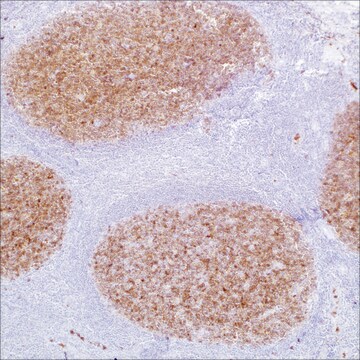
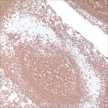
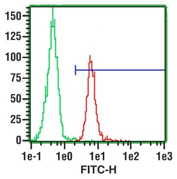
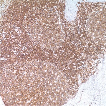
immunohistochemistry (formalin-fixed, paraffin-embedded sections): 1:25-1:100

Isotype

IgG1κ

Contrôle

tonsil

Conditions d'expédition

wet ice

Température de stockage

2-8°C

Visualisation

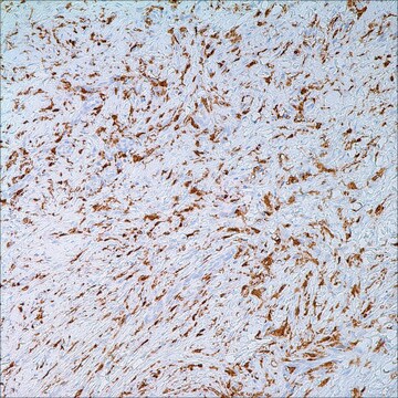
cytoplasmic, membranous

Informations sur le gène

human ... PECAM1(5175)

Description générale

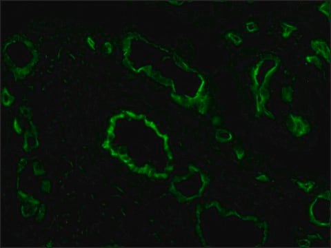
CD31, or platelet/endothelial cell adhesion molecule-1 (PECAM-1), is a vascular cell adhesionand signaling molecule that plays a role in maintaining the vascular permeability barrierat endothelial cell junctions. Immunohistochemical detection of CD31 shows cytoplasmicand membranous expression in non-neoplastic and neoplastic vascular endothelial cells,including neoplasms of vascular origin such as hemangiomas and angiosarcomas.

Liaison

CD31 Positive Control Slides, Product No. 131S, are available for immunohistochemistry (formalin-fixed, paraffin-embedded sections).

Forme physique

Solution in Tris Buffer, pH 7.3-7.7, with 1% BSA and <0.1% Sodium Azide

Notes préparatoires

Download the IFU specific to your product lot and formatNote: This requires a keycode which can be found on your packaging or product label.

Autres remarques

For Technical Service please contact: 800-665-7284 or email: service@cellmarque.com

Informations légales

Cell Marque is a trademark of Merck KGaA, Darmstadt, Germany

Vous ne trouvez pas le bon produit ?  

Essayez notre Outil de sélection de produits.

Code de la classe de stockage

12 - Non Combustible Liquids

Classe de danger pour l'eau (WGK)

WGK 2

Point d'éclair (°F)

Not applicable

Point d'éclair (°C)

Not applicable


Faites votre choix parmi les versions les plus récentes :

Certificats d'analyse (COA)

Lot/Batch Number

Vous ne trouvez pas la bonne version ?

Si vous avez besoin d'une version particulière, vous pouvez rechercher un certificat spécifique par le numéro de lot.

Déjà en possession de ce produit ?

Retrouvez la documentation relative aux produits que vous avez récemment achetés dans la Bibliothèque de documents.

Consulter la Bibliothèque de documents

R L Attanoos et al.
Thorax, 55(10), 860-863 (2000-09-19)
Three cases of diffuse malignant vascular tumours of the pleura are described which mimicked malignant mesothelioma clinically and pathologically (so called "pseudomesothelioma"). All had occupational histories of exposure to asbestos. The relationship of these tumours to mesothelioma and asbestos exposure
Malak Abedalthagafi et al.
Annals of diagnostic pathology, 14(1), 15-22 (2010-02-04)
Cutaneous angiosarcoma (AS) is a rare malignant neoplasm of dermis composed of infiltrating cells of endothelial phenotype with overall poor prognosis. Although autocrine stimulation by vascular endothelial growth factor secretion may play a role in the pathogenesis of angiosarcoma, its
Xiu-Lian Xu et al.
The American Journal of dermatopathology, 32(8), 837-840 (2010-10-01)
Microvenular hemangioma (MVH) is an uncommon benign vascular neoplasm that usually occurs as a solitary asymptomatic red or purple papule, nodule, or plaque with a predilection for the upper extremities. Patients with more than 1 lesion, that is, multiple MVHs
CD31: An Immunospecific Marker for Endothelial Differentiation in Human Neoplasms
DeYoung BR, et al.
Appl. Immunohistochem., 1, 97-100 (1993)
F Alexander-Sefre et al.
Journal of clinical pathology, 56(10), 786-788 (2003-09-30)
Lymphovascular space invasion (LVSI) is an important step in the complex process of tumour metastasis. Various methods have been used in the past to improve the histological detection of LVSI. To develop a sensitive immunohistochemical method for the detection of

Notre équipe de scientifiques dispose d'une expérience dans tous les secteurs de la recherche, notamment en sciences de la vie, science des matériaux, synthèse chimique, chromatographie, analyse et dans de nombreux autres domaines..

Contacter notre Service technique