Accéder au contenu
Merck
Toutes les photos(1)

Principaux documents

MABT819

Sigma-Aldrich

Anti-Glycosaminoglycan Antibody, skin specific Antibody, clone PG-4

clone PG-4, from mouse

Synonyme(s) :

Proteoglycan, Dermatan sulfate, Chondroitin sulfate

Se connecterpour consulter vos tarifs contractuels et ceux de votre entreprise/organisme


About This Item

Code UNSPSC :
12352203
eCl@ss :
32160702
Nomenclature NACRES :
NA.41

Source biologique

mouse

Niveau de qualité

Forme d'anticorps

purified immunoglobulin

Type de produit anticorps

primary antibodies

Clone

PG-4, monoclonal

Espèces réactives

human, chicken, shark

Conditionnement

antibody small pack of 25 μL

Technique(s)

ELISA: suitable
immunohistochemistry: suitable (paraffin)
western blot: suitable

Isotype

IgMκ

Conditions d'expédition

ambient

Modification post-traductionnelle de la cible

unmodified

Informations sur le gène

human ... FAM20B(9917)

Description générale

Proteoglycans (PG), present in all tissues, are one of the best-studied classes of matrix molecules. Over 50 members of PG have been characterized and studied for their biological significance. PG consist of a protein core to which glycosaminoglycan chains are covalently linked. Glycosaminoglycans (GAGs) are natural heteropolysaccharides that are present in every mammalian tissue. They are composed of repeating disaccharide units that consist of either sulfated or non-sulfated monosaccharides. Their molecular size and the sulfation type vary based on tissue type. The major types of GAGs found in mammalian tissues are: hyaluronan, chondroitin sulfate, and dermatan sulfate, heparin and heparan sulfate, and keratan sulfate. GAG chains are covalently bound to serine residues of the PG protein core via a tetrasaccharide linkage, consisting of xylose, two galactose residues and glucuronic acid. This antibody, clone PG-4, recognizes antigen that is associated with extracellular matrices and surfaces of cultured human dermal fibroblasts and the surfaces of cultured human adult keratinocytes. This antibody recognize native epitopes located within glycosaminoglycan chains and is detects both chondroitin sulfate (Versican) and dermatan sulfate (Decorin and Biglycan) proteoglycans. Treatment of PG with chondroitinases is reported to abolish its immunoreactivity for all antigens. (Ref.: Afratis, N et al. (2012). FEBS Journal 279 (7); 1177-1197; Sorrell, JM et al. (1999). Histochem. J. 31 (8); 549-558).

Spécificité

Clone PG4 detects glycosaminoglycan in skin cells.

Immunogène

Crude membrane fractions from adult human dermal fibroblasts from breast skin.

Application

Anti-Glycosaminoglycan, skin specific, clone PG-4, Cat. No. MABT819, is a mouse monoclonal antibody that detects glycosaminoglycan and has been tested for use in ELISA, Immunohistochemistry (Paraffin), and Western Blotting.
Research Category
Cell Structure
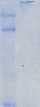
Western Blotting Analysis: A representative lot detected Glycosaminoglycan in Western Blotting applications (Sorrell, J.M., et. al. (1999). Histochem J. 31(8):549-58).

ELISA Analysis: A representative lot detected Glycosaminoglycan in ELISA applications (Sorrell, J.M., et. al. (1999). Histochem J. 31(8):549-58).

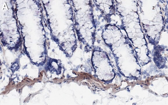
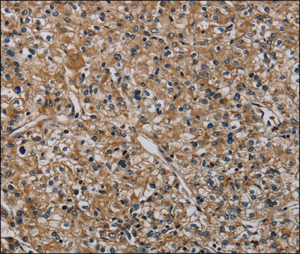
Immunohistochemistry Analysis: A representative lot detected Glycosaminoglycan in Immunohistochemistry applications (Sorrell, J.M., et. al. (1999). Histochem J. 31(8):549-58).

Qualité

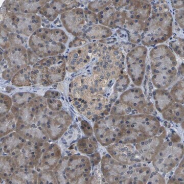
Evaluated by Immunohistochemistry (Paraffin) in human skin tissue.

Immunohistochemistry Analysis: A 1:250 dilution of this antibody detected Glycosaminoglycan in human skin tissue.

Description de la cible

~60 kDa observed. Uncharacterized bands may be observed in some lysate(s).

Forme physique

Format: Purified
Protein G purified
Purified mouse monoclonal antibody IgM in buffer containing 0.1 M Tris-Glycine (pH 7.4), 150 mM NaCl with 0.05% sodium azide.

Stockage et stabilité

Stable for 1 year at 2-8°C from date of receipt.

Autres remarques

Concentration: Please refer to lot specific datasheet.

Clause de non-responsabilité

Unless otherwise stated in our catalog or other company documentation accompanying the product(s), our products are intended for research use only and are not to be used for any other purpose, which includes but is not limited to, unauthorized commercial uses, in vitro diagnostic uses, ex vivo or in vivo therapeutic uses or any type of consumption or application to humans or animals.

Vous ne trouvez pas le bon produit ?  

Essayez notre Outil de sélection de produits.

Code de la classe de stockage

12 - Non Combustible Liquids

Classe de danger pour l'eau (WGK)

WGK 1

Point d'éclair (°F)

does not flash

Point d'éclair (°C)

does not flash


Certificats d'analyse (COA)

Recherchez un Certificats d'analyse (COA) en saisissant le numéro de lot du produit. Les numéros de lot figurent sur l'étiquette du produit après les mots "Lot" ou "Batch".

Déjà en possession de ce produit ?

Retrouvez la documentation relative aux produits que vous avez récemment achetés dans la Bibliothèque de documents.

Consulter la Bibliothèque de documents

Mahasish Shome et al.
Methods in molecular biology (Clifton, N.J.), 2597, 131-142 (2022-11-15)
Protein microarrays are an important tool when analyzing multiple analytes simultaneously. As the human genome contains approximately 20,000 genes, examining the interactions of even just one representative protein for each gene requires a high-throughput technique. For instance, the interaction between

Notre équipe de scientifiques dispose d'une expérience dans tous les secteurs de la recherche, notamment en sciences de la vie, science des matériaux, synthèse chimique, chromatographie, analyse et dans de nombreux autres domaines..

Contacter notre Service technique