Accéder au contenu
Merck
Toutes les photos(3)

Principaux documents

MABT326

Sigma-Aldrich

Anti-CEACAM3/5 Antibody, clone 308/3-3

clone 308/3-3, from mouse

Synonyme(s) :

Carcinoembryonic antigen-related cell adhesion molecule 3, Carcinoembryonic antigen CGM1, CD66d, Carcinoembryonic antigen-related cell adhesion molecule 5, Carcinoembryonic antigen, CEA, Meconium antigen 100, CD66e

Se connecterpour consulter vos tarifs contractuels et ceux de votre entreprise/organisme


About This Item

Code UNSPSC :
12352203
eCl@ss :
32160702
Nomenclature NACRES :
NA.43
Clone:
308/3-3, monoclonal
application:
ELISA
FACS
IP
WB
Espèces réactives:
human
Technique(s):
ELISA: suitable
flow cytometry: suitable
immunoprecipitation (IP): suitable
western blot: suitable
citations:

Source biologique

mouse

Niveau de qualité

Forme d'anticorps

purified antibody

Type de produit anticorps

primary antibodies

Clone

308/3-3, monoclonal

Espèces réactives

human

Ne doit pas réagir avec

rat, mouse

Technique(s)

ELISA: suitable
flow cytometry: suitable
immunoprecipitation (IP): suitable
western blot: suitable

Isotype

IgG1κ

Numéro d'accès NCBI

Numéro d'accès UniProt

Modification post-traductionnelle de la cible

unmodified

Informations sur le gène

human ... CEACAM5(1048)

Description générale

Carcinoembryonic antigen-related cell adhesion molecule 3 (UniProt P40198; also known as Carcinoembryonic antigen CGM1, CD66d) is encoded by the CEACAM3 (also known as CD66D, CGM1) gene (Gene ID 1084) in human. The carcinoembryonic antigen- (CEA-) related cell adhesion molecules (CEACAMs) constitute a 12-member subgroup of the CEA family of immunoglobulin-related proteins first described in 1965 (PMID 4953873). CEACAMs are reported to participate in diverse physiological processes, including cell adhesion, differentiation, proliferation, and survival, as well as in carcinogenesis and bacterial pathogenesis. CEACAM molecules from neighbouring cells can interact via their respective extracellular N-terminal IgV-like domain and mediate cell-to-cell adhesion through trans-oligomerization. CEACAM molecules within the same cell can also undergo transmembrane domain-mediated cis-oligomerization, an event important for sustaining downstream cellular signaling. Athough CEACAM1 is shown to utilize its cytoplasmic domain for transducing cellular signaling, not all CEACAM members are expressed with a significant cytoplasmic domain, and others (CEACAM5/6/7/8) are GPI-anchored without even a transmembrane domain. Human CEACAM3 and CEACAM4 are two CEACAMs whose extracellular region is composed of only one V-type Ig-like extracellular domain (a.a. 35-142 of CEACAM3). Human CEACAM5 (UniProt P06731), on the other hand, is a GPI-anchored CEACAM with the most extracellular Ig domains, 7 in all.

Spécificité

Clone 308/3-3 reacts with human CEACAM3 and CEACAM5, but not human CEACAM1/4/6/7/8, rat CEACAM1, or mouse CEACAM1 & CEACAM2.

Immunogène

Epitope: Extracellular domain.
Human IgG1 Fc-tagged recombinant human CEACAM3 extracellular domain.

Application

Anti-CEACAM Antibody, clone 308/3-3 is an antibody against CEACAM3/5 for use in Western Blotting, ELISA, Flow Cytometry, Immunoprecipitation.
ELISA Analysis: 10 μg/mL from a representative lot detected CHO cell-expressed full-length glycosylated human CEACAM3 and CEACAM5, but not CEACAM1 (CEACAM1-4L), CEACAM4, CEACAM6, CEACAM7, or CEACAM8 (Courtesy of Dr. Bernhard B. Singer, University of Duisburg-Essen, Germany).
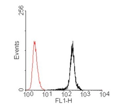
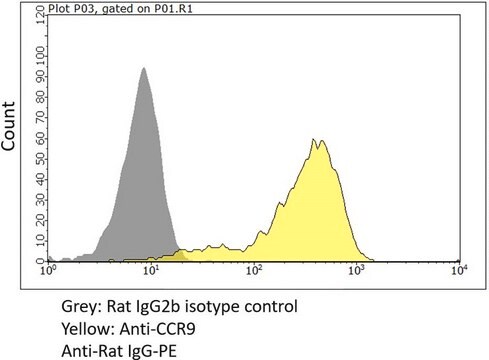
Flow Cytometry Analysis: 10 μg/mL from a representative lot immunostained CHO cell transfectants expressing human CEACAM3 and CEACAM5, but not CHO cells expressing human CEACAM1 (CEACAM1-4L), CEACAM4, CEACAM6, CEACAM7, or CEACAM8 (Courtesy of Dr. Bernhard B. Singer, University of Duisburg-Essen, Germany).
Immunoprecipitation Analysis: 5 μg from a representative lot immunoprecipitated exogenously expressed full-length glycosylated human CEACAM5 from 500 μg of lysate (in 600 μL) from transfected HeLa cells (Courtesy of Dr. Bernhard B. Singer, University of Duisburg-Essen, Germany).
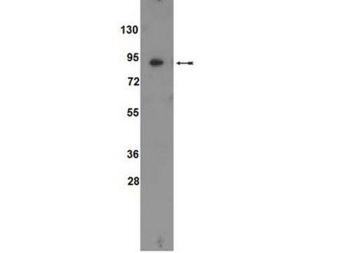
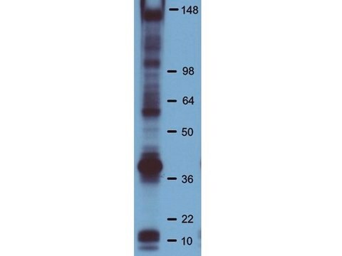
Western Blotting Analysis: 10 μg/mL from a representative lot detected exogenously expressed full-length glycosylated human CEACAM5 in 50 μg of lysate from transfected HeLa cells (Courtesy of Dr. Bernhard B. Singer, University of Duisburg-Essen, Germany).
Research Category
Cell Structure
Research Sub Category
Adhesion (CAMs)

Qualité

Evaluated by Western Blotting Analysis in lysates of CEACAM3 & CEACAM5 transfected CHO cells.

Western Blotting Analysis: 2.0 μg/mL of this antibody detected exogenously expressed full-length glycosylated human CEACAM3 and CEACAM5 in 10 μg of lysates from transfected CHO cells.

Description de la cible

~30/180 kDa (CEACAM3/CEACAM5) observed. 27.09/19.40/22.75 kDa (human CEACAM3 isoform CGM1A/CGM1B/CGM1C) and 76.80/76.72 kDa (human CEACAM5 isoform 1/2) calculated. Target bands appear smeary and larger than the calculated molecular weights due to glycosylation. Uncharacterized band(s) may appear in some lysates.

Forme physique

Format: Purified
Protein G Purified
Purified mouse monoclonal IgG1κ in buffer containing 0.1 M Tris-Glycine (pH 7.4), 150 mM NaCl with 0.05% sodium azide.

Stockage et stabilité

Stable for 1 year at 2-8°C from date of receipt.

Autres remarques

Concentration: Please refer to lot specific datasheet.

Clause de non-responsabilité

Unless otherwise stated in our catalog or other company documentation accompanying the product(s), our products are intended for research use only and are not to be used for any other purpose, which includes but is not limited to, unauthorized commercial uses, in vitro diagnostic uses, ex vivo or in vivo therapeutic uses or any type of consumption or application to humans or animals.

Vous ne trouvez pas le bon produit ?  

Essayez notre Outil de sélection de produits.

Code de la classe de stockage

12 - Non Combustible Liquids

Classe de danger pour l'eau (WGK)

WGK 1

Point d'éclair (°F)

Not applicable

Point d'éclair (°C)

Not applicable


Certificats d'analyse (COA)

Recherchez un Certificats d'analyse (COA) en saisissant le numéro de lot du produit. Les numéros de lot figurent sur l'étiquette du produit après les mots "Lot" ou "Batch".

Déjà en possession de ce produit ?

Retrouvez la documentation relative aux produits que vous avez récemment achetés dans la Bibliothèque de documents.

Consulter la Bibliothèque de documents

Notre équipe de scientifiques dispose d'une expérience dans tous les secteurs de la recherche, notamment en sciences de la vie, science des matériaux, synthèse chimique, chromatographie, analyse et dans de nombreux autres domaines..

Contacter notre Service technique