Accéder au contenu
Merck
Toutes les photos(1)

Key Documents

MABF939

Sigma-Aldrich

Anti-IFNAR1 Antibody, clone 4G8

clone 4G8, from mouse

Synonyme(s) :

Interferon alpha/beta receptor 1, CRF2-1, Cytokine receptor class-II member 1, Cytokine receptor family 2 member 1, IFN-alpha/beta receptor 1, IFN-R-1, Type I interferon receptor 1

Se connecterpour consulter vos tarifs contractuels et ceux de votre entreprise/organisme


About This Item

Code UNSPSC :
12352203
eCl@ss :
32160702

Source biologique

mouse

Niveau de qualité

Forme d'anticorps

purified immunoglobulin

Type de produit anticorps

primary antibodies

Clone

4G8, monoclonal

Espèces réactives

human

Technique(s)

flow cytometry: suitable

Isotype

IgG1κ

Numéro d'accès NCBI

Numéro d'accès UniProt

Conditions d'expédition

ambient

Modification post-traductionnelle de la cible

unmodified

Informations sur le gène

human ... IFNAR1(3454)

Description générale

Interferon alpha/beta receptor 1 (UniProt P17181; also known as CRF2-1, Cytokine receptor class-II member 1, Cytokine receptor family 2 member 1, IFN-alpha/beta receptor 1, IFN-R-1, Type I interferon receptor 1) is encoded by the IFNAR1 (also known as IFNAR) gene (Gene ID 3454) in human. Interferons (IFNs) are cytokines whose prouction and release by host cells are induced in the presence of pathogens (e.g. viruses, bacteria, parasites) and tumor cells. More than twenty distinct IFN genes and proteins have been identified and classified into three main types. Type I IFNs (IFN-α, IFN-β, IFN-ε, IFN-κ, and IFN-ω) exert their function via binding IFN-α/β receptor (encoded by the IFNAR1 and IFNAR2 gene, respectively). Ligation of IFNAR by IFN-I results in activation of the Janus kinases, Jak1 and Tyk2, which in turn phosphorylates STAT1 and STAT2, resulting in recruitment of IRF9 to form the IFN-stimulated gene factor 3 (ISGF3) transcription factor. ISGF3 then translocates to the nucleus, where it binds IFN-stimulated response elements (ISREs) and drives the transcription of IFN-stimulated genes (ISGs). IFNAR1 is phosphorylated by Tyk2 and PKD2 upon IFN-I stimulation, leading to IFNAR1 ubiquitination and lysosomal degradation. IFNAR1 is also
phosphorylated by p38 MAP kinase in response to non-IFN stimuli, including the PERK-dependent unfolded protein response (UPR), ligation of pattern recognition receptors (PRRs), or through signaling via other inflammatory cytokines or growth factors including VEGF, IL-1β and TNFα. Encephalitic flaviviruses antagonize IFN-I signaling by inhibiting IFNAR1 surface expression, where the viral nonstructural protein 5 (NS5) targets cellular prolidase (PEPD) that is required for IFNAR1 maturation and accumulation.

Spécificité

Clone 4G8 detected a loss of HEK293 cell surface IFNAR1 immunoreactivity following lentivirus-mediated cellular IFNAR1 shRNA delivery or West Nile virus (WNV) infection (Lubick, K.J., et al. (2015). Cell Host Microbe. 18(1):61-74).

Immunogène

Epitope: extracellular domain
His-tagged recombinant human IFNAR1 extracellular domain.

Application

Flow Cytometry Analysis: A representative lot detected a time-dependent loss of HEK293 cell surface IFNAR1 immunoreactivity following West Nile virus (WNV) infection. A significantly faster surface IFNAR1 loss was observed when cells were infected with WNV that produces IFN-I-antagonizing NS5 (nonstructural protein 5) with S653F mutation (Lubick, K.J., et al. (2015). Cell Host Microbe. 18(1):61-74).

Flow Cytometry Analysis: A representative lot detected a loss of HEK293 cell surface IFNAR1 immunoreactivity following lentivirus-mediated cellular IFNAR1 shRNA delivery (Lubick, K.J., et al. (2015). Cell Host Microbe. 18(1):61-74).
Research Category
Inflammation & Immunology
This mouse monoclonal Anti-IFNAR1 Antibody, clone 4G8, Cat. No. MABF939 is validated for use in Flow Cytometry for the detection of IFNAR1.

Qualité

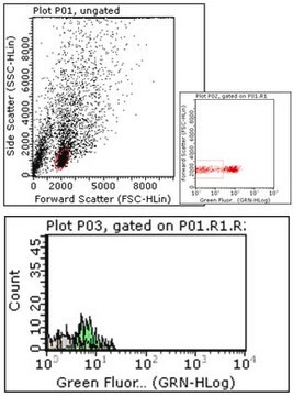
Evaluated by Flow Cytometry in K562 cells.

Flow Cytometry Analysis: 1 µg of this antibody detected IFNAR1 on the surface of K562 cells.

Description de la cible

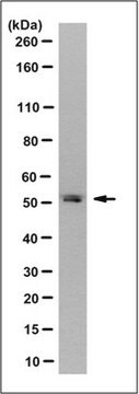
63.53/60.93 kDa (pro-/mature isoform 1), 49.55/46.95 kDa (pro-/mature isoform 2), 56.72/54.12 kDa (pro-/mature isoform 3), 56.05 kDa (isoform 4) calculated. Apparent molecular weight appears greater than 100 kDa due to glycosylation.

Forme physique

Format: Purified
Protein G purified.
Purified mouse IgG1κ in buffer containing 0.1 M Tris-Glycine (pH 7.4), 150 mM NaCl with 0.05% sodium azide.

Stockage et stabilité

Stable for 1 year at 2-8°C from date of receipt.

Autres remarques

Concentration: Please refer to lot specific datasheet.

Clause de non-responsabilité

Unless otherwise stated in our catalog or other company documentation accompanying the product(s), our products are intended for research use only and are not to be used for any other purpose, which includes but is not limited to, unauthorized commercial uses, in vitro diagnostic uses, ex vivo or in vivo therapeutic uses or any type of consumption or application to humans or animals.

Vous ne trouvez pas le bon produit ?  

Essayez notre Outil de sélection de produits.

Code de la classe de stockage

12 - Non Combustible Liquids

Classe de danger pour l'eau (WGK)

WGK 1

Point d'éclair (°F)

Not applicable

Point d'éclair (°C)

Not applicable


Certificats d'analyse (COA)

Recherchez un Certificats d'analyse (COA) en saisissant le numéro de lot du produit. Les numéros de lot figurent sur l'étiquette du produit après les mots "Lot" ou "Batch".

Déjà en possession de ce produit ?

Retrouvez la documentation relative aux produits que vous avez récemment achetés dans la Bibliothèque de documents.

Consulter la Bibliothèque de documents

Notre équipe de scientifiques dispose d'une expérience dans tous les secteurs de la recherche, notamment en sciences de la vie, science des matériaux, synthèse chimique, chromatographie, analyse et dans de nombreux autres domaines..

Contacter notre Service technique