ABT119
Anti-CELSR1 Antibody
from rabbit, purified by affinity chromatography
Synonyme(s) :
Cadherin EGF LAG seven-pass G-type receptor 1, Cadherin family member 9, Flamingo homolog 2, hFmi2
About This Item
Produits recommandés
Source biologique
rabbit
Niveau de qualité
Forme d'anticorps
affinity isolated antibody
Type de produit anticorps
primary antibodies
Clone
polyclonal
Produit purifié par
affinity chromatography
Espèces réactives
rat
Réactivité de l'espèce (prédite par homologie)
mouse (based on 100% sequence homology), human (based on 100% sequence homology)
Technique(s)
immunofluorescence: suitable
immunohistochemistry: suitable
western blot: suitable
Numéro d'accès NCBI
Numéro d'accès UniProt
Conditions d'expédition
wet ice
Modification post-traductionnelle de la cible
unmodified
Informations sur le gène
human ... CELSR1(9620)
Description générale
Immunogène
Application
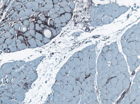
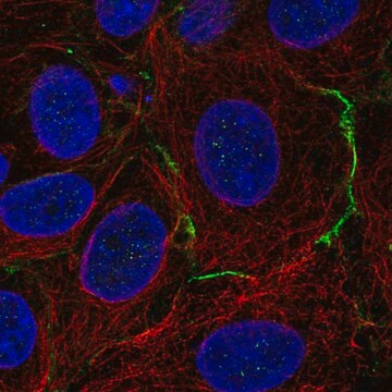
Immunohistochemistry Analysis: A 1:500 dilution from a representative lot detected CELSR1 in neurons of rat cortex tissue and in Purkinje cells and cells within the granular layer of rat cerebellum tissue.
Qualité
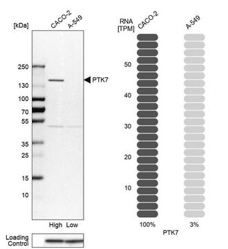
Western Blot Analysis: 1 µg/mL of this antibody detected CELSR1 in 10 µg of PC12 cell lysate.
Description de la cible
An uncharacterized band at ~54 kDa may be observed in some cell lysates.
Autres remarques
Vous ne trouvez pas le bon produit ?
Essayez notre Outil de sélection de produits.
Code de la classe de stockage
12 - Non Combustible Liquids
Classe de danger pour l'eau (WGK)
WGK 1
Point d'éclair (°F)
Not applicable
Point d'éclair (°C)
Not applicable
Certificats d'analyse (COA)
Recherchez un Certificats d'analyse (COA) en saisissant le numéro de lot du produit. Les numéros de lot figurent sur l'étiquette du produit après les mots "Lot" ou "Batch".
Déjà en possession de ce produit ?
Retrouvez la documentation relative aux produits que vous avez récemment achetés dans la Bibliothèque de documents.
Notre équipe de scientifiques dispose d'une expérience dans tous les secteurs de la recherche, notamment en sciences de la vie, science des matériaux, synthèse chimique, chromatographie, analyse et dans de nombreux autres domaines..
Contacter notre Service technique