Accéder au contenu
Merck
Toutes les photos(3)

Principaux documents

07-1483

Sigma-Aldrich

Anti-IκBα Antibody

serum, from rabbit

Se connecterpour consulter vos tarifs contractuels et ceux de votre entreprise/organisme


About This Item

Code UNSPSC :
12352203
eCl@ss :
32160702
Nomenclature NACRES :
NA.41

Source biologique

rabbit

Niveau de qualité

Forme d'anticorps

serum

Type de produit anticorps

primary antibodies

Clone

polyclonal

Espèces réactives

rat, mouse, human

Technique(s)

ELISA: suitable
immunohistochemistry: suitable (paraffin)
immunoprecipitation (IP): suitable
western blot: suitable

Isotype

IgG

Numéro d'accès NCBI

Numéro d'accès UniProt

Conditions d'expédition

dry ice

Modification post-traductionnelle de la cible

unmodified

Informations sur le gène

human ... NFKBIA(4792)

Description générale

IKK beta (IκB Kinase, IKKβ, I-Kappa-B kinase-beta) is a member of the IKK complex which is composed of IKK alpha, IKK beta, IKK gamma and IKAP. IκB Kinase (IKK) complex is a critical component in NFκB regulation. In a majority of unstimulated cells, the NFκB transcription factors exist in their inactive form and are retained in the cytoplasm by the bound inhibitory IκB proteins. For NFκB to become activated, it must first disassociate from the inhibitor IκB. This occurs via the serine phosphorylation of IκB on Ser32 and Ser36. Post receptor stimulation, most notably by TNFα, NIK, a serine/threonine kinase, activates and phosphorylates IKKα and IKKβ. Phosphorylation of 2 sites at the activation loop of IKK beta is essential for activation of IKK by TNF and IL1. Once activated, IKK beta autophosphorylates, which in turn decreases IKK activity and prevents prolonged activation of the inflammatory response. Additionally, IKK beta activity can also be regulated by MEKK-1. IKK then binds to and phosphorylates IκBα on the two serine residues. This phosphorylation marks IκB for ubiquination and destruction by the proteosome. This frees the NFκB complex and allows it to translocate to the nucleus.

Spécificité

Recognizes IκBα, C-terminus. May react non-specifically with other proteins. Control peptide provided will compete only with the specific reaction of antiserum with IκBα.

Immunogène

Epitope: C-terminus
IκBα peptide corresponding to a region near the C-terminus of the human protein, conjugated to KLH.

Application

Detect IκBα using this Anti-IκBα Antibody validated for use in WB, IH(P), ELISA & IP.
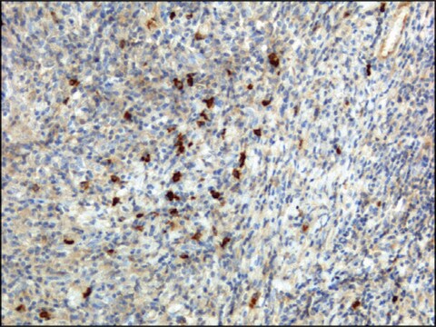
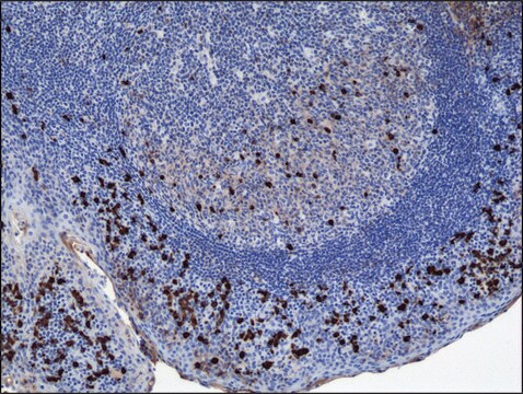
Immunohistochemisty (paraffin): Representative testing from a previous lot.
Anti-IкBα staining on invasive ductal carcinoma tissue (Breast Cancer) was pretreated with citrate buffer, pH 6.0. A 1:1,000 diluted was used using IHC-Select detection with HRP-DAB.


ELISA: Recommended

Immunoprecipitation: Recommended

Optimal dilutions must be determined by the end user.
Research Category
Epigenetics & Nuclear Function
Research Sub Category
Transcription Factors

Qualité

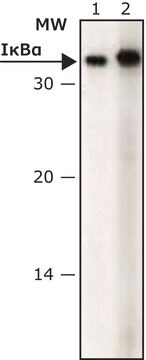
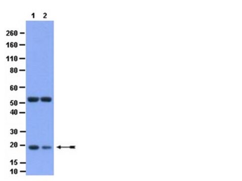
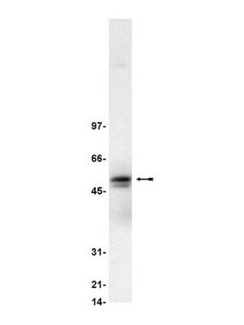
Routinely evaluated by Western Blot on HEK293 lysates.

Western Blot Analysis:
1:500 to 1:2,000 dilution of this lot detected IkBalpha on 10 μg of HEK293 lysates.

Description de la cible

~36 kDa

Liaison

Replaces: AB3016

Forme physique

Rabbit polyclonal IgG serum in buffer containing 0.02 M Potassium phosphate with 0.15 M NaCl, pH 7.2 and 0.1% sodium azide.

Stockage et stabilité

Both antibody and control are stable for 6 months at -20ºC in undiluted aliquots from date of receipt.
Handling Recommendations: Upon first thaw, and prior to removing the cap, centrifuge the vial and gently mix the solution. Aliquot into microcentrifuge tubes and store at -20°C. Avoid repeated freeze/thaw cycles, which may damage IgG and affect product performance.

Remarque sur l'analyse

Control
HEK293 cell lysate.
Control Peptide: Included with the antibody is 50 μg (1 mg/mL) of IκBα control peptide. The peptide will block the specific interaction of AB3016 with the IκBα subunit. Control peptide should be used at 1.0 μg per 1.0 μL of antiserum used in assay. Optimal concentrations must be determined by the end user.

Clause de non-responsabilité

Unless otherwise stated in our catalog or other company documentation accompanying the product(s), our products are intended for research use only and are not to be used for any other purpose, which includes but is not limited to, unauthorized commercial uses, in vitro diagnostic uses, ex vivo or in vivo therapeutic uses or any type of consumption or application to humans or animals.

Vous ne trouvez pas le bon produit ?  

Essayez notre Outil de sélection de produits.

Code de la classe de stockage

12 - Non Combustible Liquids

Classe de danger pour l'eau (WGK)

WGK 2

Point d'éclair (°F)

Not applicable

Point d'éclair (°C)

Not applicable


Certificats d'analyse (COA)

Recherchez un Certificats d'analyse (COA) en saisissant le numéro de lot du produit. Les numéros de lot figurent sur l'étiquette du produit après les mots "Lot" ou "Batch".

Déjà en possession de ce produit ?

Retrouvez la documentation relative aux produits que vous avez récemment achetés dans la Bibliothèque de documents.

Consulter la Bibliothèque de documents

Yu-Chen Hou et al.
American journal of physiology. Lung cellular and molecular physiology, 302(1), L174-L183 (2011-10-18)
Lung epithelial cells are important barriers in the respiratory system that provoke inflammatory responses through nuclear factor (NF)-κB activation to prevent pathogens from invading the body. Lipopolysaccharide (LPS) is a common pathogen-associated stimulus that activates IκB kinase (IKK) to regulate
Bhargab Kalita et al.
PloS one, 11(12), e0168525-e0168525 (2016-12-31)
Development of an effective radio protector to minimise radiation-inflicted damages have largely failed owing to inherent toxicity of most of the agents examined so far. This study is centred towards delivering protection to lethally irradiated mice by pre-administration of a

Notre équipe de scientifiques dispose d'une expérience dans tous les secteurs de la recherche, notamment en sciences de la vie, science des matériaux, synthèse chimique, chromatographie, analyse et dans de nombreux autres domaines..

Contacter notre Service technique