Skip to Content
Merck
All Photos(6)

Key Documents

M8421

Sigma-Aldrich

Monoclonal Anti-Myosin (Skeletal, Slow) antibody produced in mouse

clone NOQ7.5.4D, ascites fluid

Synonym(s):

Anti-CMD1S, Anti-CMH1, Anti-MPD1, Anti-MYHCB, Anti-SPMD, Anti-SPMM

Sign Into View Organizational & Contract Pricing


About This Item

MDL number:
UNSPSC Code:
12352203
NACRES:
NA.41

biological source

mouse

Quality Level

conjugate

unconjugated

antibody form

ascites fluid

antibody product type

primary antibodies

clone

NOQ7.5.4D, monoclonal

contains

15 mM sodium azide

species reactivity

sheep, rat, bovine, hamster, pig, canine, feline, goat, chicken, mouse, rabbit, human, guinea pig

packaging

antibody small pack of 25 μL

technique(s)

electron microscopy: suitable
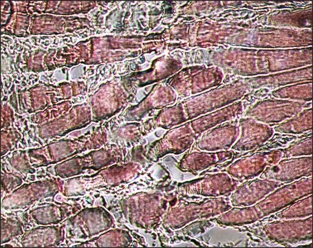
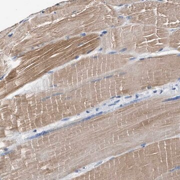
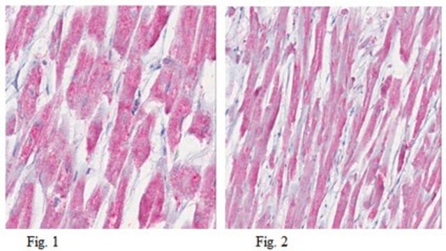
immunohistochemistry (formalin-fixed, paraffin-embedded sections): 1:4,000 using protease-digested, sections of rabbit tongue
indirect ELISA: suitable
radioimmunoassay: suitable
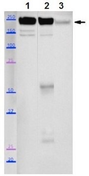
western blot: 1:5,000 using extract of rat or rabbit tongue

isotype

IgG1

UniProt accession no.

shipped in

dry ice

storage temp.

−20°C

target post-translational modification

unmodified

Gene Information

human ... MYH7(4625)
mouse ... Myh7(140781)
rat ... Myh7(29557)

General description

Myosin is a 480 kDa protein known to interact with actin in muscle and in non-muscle cells. It contains two identical heavy chains (200 kDa each) and four light chains (15-26 kDa). Myosin 7 (MYH7) is mapped to human chromosome 14q11.1.
Monoclonal Anti-Myosin (Skeletal, Slow) (mouse IgG1 isotype) is derived from the NOQ7.5.4D hybridoma produced by the fusion of mouse myeloma cells and splenocytes from BALB/c mice. Myosin, purified from myofibrils isolated from human skeletal muscle, was used as the immunogen.1-3. The isotype is determined by a double diffusion immunoassay using Mouse Monoclonal Antibody Isotyping Reagents, Catalog Number ISO2.

Specificity

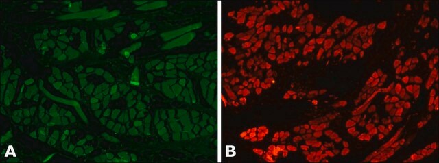
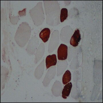
Monoclonal Anti-Myosin (Skeletal, Slow) recognizes an epitope located on the heavy meromyosin portion of human adult skeletal muscle slow myosin. Cross-reactivity has been observed with human, bovine, sheep, goat, porcine, dog, cat, rabbit, hamster, guinea pig, rat, mouse, and chicken. In rat, the product is highly specific for the slow myosin heavy chain of adult skeletal muscle. It does not react with the fast myosin heavy chain. It does not cross-react with embryonic or neonatal rat skeletal myosins, nor with cat fetal/embryonic skeletal myosin. Using immunohistochemical staining, the antibody reacts with human adult type I skeletal muscle fibers and with cardiac muscle cells. It also reacts faintly with adult human C-Fibers, co-expressing myosin heavy chain I and IIa in variable ratios. The product shows no reactivity with human or rat adult smooth muscle or with non-muscle tissues.
Reacts with human adult skeletal muscle slow myosin. Does not react with the fast myosin heavy chain nor with embryonic or neonatal rat skeletal myosins, nor with cat fetal/embryonic skeletal myosin. Also reacts with human adult type I skeletal muscle fibers and with cardiac muscle cells. No reactivity detected with human or rat adult smooth muscle or with non-muscle tissues.

Immunogen

Human skeletal muscle myosin purified from myofibrils.

Application

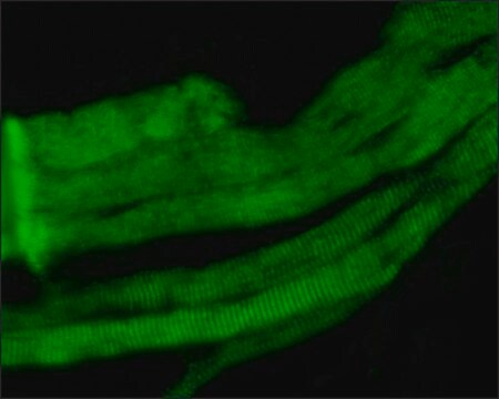
Monoclonal Anti-Myosin (Skeletal, Slow) may be used for the localization of slow (Type I) myosin.
Monoclonal Anti-Myosin (Skeletal, Slow) antibody has been used in the detection of Myosin 7 using:
  • light microscopy
  • immunofluorescence staining
  • immunoblotting
  • ELISA
  • solid-phase RIA
  • immunohistology (frozen, formalin-fixed, paraffin-embedded and methacarn-fixed paraffin-embedded tissue sections)
  • immunoelectronmicroscopy
Detection of slow myosin in formalin-fixed, paraffin-embedded, tissues is greatly enhanced by proteolytic digestion of the preparation.

Biochem/physiol Actions

Myosin molecules consist of two major regions: tail (rod) and heads; they aggregate into filaments through the tail region and interact with actin and with ATP through the head region. Multiple forms of myosin heavy chains exist for each muscle type: skeletal, cardiac, smooth and in non-muscle. Myosin isoforms exist in different types of skeletal muscle, depending on the physiological function of the muscle. Mammalian muscle fibers are classified primarily into slow (I), fast-red (IIa) and fast-white (IIb) major types. Changes in the speed of muscle contraction brought about by neural influences result from changes in the pattern of expression of myosin and other myofibrillar genes.
Transient expression of different myosin isoforms occurs during fetal growth and development. Mutations in myosin 7 is associated with laing distal myopathy (LDM). Myosin 7 gene mutations results in muscular dystrophy diseases like scapuloperoneal myopathy. Mutations leads to storage of myosin protein aggregates in muscle, leading to myosin storage myopathy. Mutations in MYH7 is also associated with hypertrophic cardiomyopathy and in heart malformation disease called ebstein anomaly.

Physical form

Supplied as ascites fluid with 15 mM sodium azide as a preservative.

Storage and Stability

For continuous use, store at 2-8 °C for up to one month. For extended storage freeze in working aliquots. Repeated freezing and thawing, or storage in "frostfree" freezers, is not recommended. If slight turbidity occurs upon prolonged storage, clarify the solution by centrifugation before use.

Disclaimer

Unless otherwise stated in our catalog or other company documentation accompanying the product(s), our products are intended for research use only and are not to be used for any other purpose, which includes but is not limited to, unauthorized commercial uses, in vitro diagnostic uses, ex vivo or in vivo therapeutic uses or any type of consumption or application to humans or animals.

Not finding the right product?  

Try our Product Selector Tool.

Storage Class Code

12 - Non Combustible Liquids

WGK

WGK 3

Flash Point(F)

Not applicable

Flash Point(C)

Not applicable


Certificates of Analysis (COA)

Search for Certificates of Analysis (COA) by entering the products Lot/Batch Number. Lot and Batch Numbers can be found on a product’s label following the words ‘Lot’ or ‘Batch’.

Already Own This Product?

Find documentation for the products that you have recently purchased in the Document Library.

Visit the Document Library

TGFβ contributes to impaired exercise response by suppression of mitochondrial key regulators in skeletal muscle.
Bohm A, et al.
Diabetes, db151723-db151723 (2016)
Developmental myosins: expression patterns and functional significance.
Schiaffino S, et al.
Skeletal Muscle, 5(1), 22-22 (2015)
A de novo mutation of the MYH7 gene in a large Chinese family with autosomal dominant myopathy.
Oda T, et al.
Human Genome Variation, 2, 15022-15022 (2015)
Mutations in the sarcomere gene MYH7 in Ebstein anomaly.
Postma AV, et al.
Circulation: Genomic and Precision Medicine, 4(1), 43-50 (2011)
The hypertrophic cardiomyopathy myosin mutation R453C alters ATP binding and hydrolysis of human cardiac β-myosin.
Bloemink M, et al.
The Journal of Biological Chemistry, 289(8), 5158-5167 (2014)

Our team of scientists has experience in all areas of research including Life Science, Material Science, Chemical Synthesis, Chromatography, Analytical and many others.

Contact Technical Service