Direkt zum Inhalt
Merck

358R-1

Sigma-Aldrich

MUM1 (EP190) Rabbit Monoclonal Primary Antibody

Synonym(e):

Multiple Myeloma Oncogene-1

Anmeldenzur Ansicht organisationsspezifischer und vertraglich vereinbarter Preise


About This Item

UNSPSC-Code:
12352203

Biologische Quelle

rabbit

Qualitätsniveau

100
500

Konjugat

unconjugated

Antikörperform

culture supernatant

Antikörper-Produkttyp

primary antibodies

Klon

EP190, monoclonal

Beschreibung

For In Vitro Diagnostic Use in Select Regions

Form

buffered aqueous solution

Speziesreaktivität

human

Verpackung

vial of 0.1 mL concentrate (358R-14)
vial of 0.1 mL concentrate Research Use Only (358R-14-RUO)
vial of 0.5 mL concentrate (358R-15)
vial of 1.0 mL concentrate (358R-16)
vial of 1.0 mL concentrate Research Use Only (358R-16-RUO)
vial of 1.0 mL pre-dilute Research Use Only (358R-17-RUO)
vial of 1.0 mL pre-dilute ready-to-use (358R-17)
vial of 7.0 mL pre-dilute ready-to-use (358R-18)
vial of 7.0 mL pre-dilute ready-to-use Research Use Only (358R-18-RUO)

Hersteller/Markenname

Cell Marque

Methode(n)

immunohistochemistry (formalin-fixed, paraffin-embedded sections): 1:100-1:500 (concentrated)

Isotyp

IgG

Kontrolle

diffuse large B-cell (lymphoma), plasma cell myeloma (tumor), tonsil

Versandbedingung

wet ice

Lagertemp.

2-8°C

Visualisierung

cytoplasmic, nuclear

Angaben zum Gen

human ... PWWP3A(84939)

Allgemeine Beschreibung

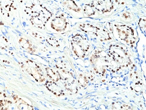
Anti-MUM1 antibody labels a 50kDa, multiple myeloma oncogene-1 (MUM1) protein. MUM1 is encoded by the MUM1/IRF-4 gene, which is mapped to 6q23-25 and identified as a myeloma-associated oncogene. It is a member of the interferon regulatory factor family of transcription factors and plays an important role in the regulation of gene expression in response to signaling by interferon and other cytokines. MUM1 positive cells express the protein in the nucleus in a diffuse and microgranular pattern. However, some positivity is also observed in the cytoplasm of MUM1-expressing cells. In normal/reactive lymphoid tissues, such as lymph node, this antibody stains plasma cells, some B-cells in the light zone of germinal centers, and a subset of T-cells (T-cells in germinal centers and interfollicular areas). MUM1 expression has been described in diffuse large B-cell lymphoma (DLBCL). Anti-MUM1 antibody can stain other B-cell lymphomas such as lymphoplasmacytic lymphoma, chronic lymphocytic leukemia, follicular lymphoma, marginal zone lymphoma, lymphoblastic lymphoma/leukemia, primary effusion lymphoma, DLBCL, Burkitt-like lymphoma, and classical Hodgkin lymphoma. However, the tumor cells in nodular lymphocyte predominant Hodgkin lymphoma are negative or only weakly positive. MUM1 is also expressed in plasma cell myeloma.

Qualität


IVD

IVD

IVD

RUO

Verlinkung

MUM1 Positive Control Slides, Product No. 358S, are available for immunohistochemistry (formalin-fixed, paraffin-embedded sections).

Physikalische Form

Solution in Tris Buffer, pH 7.3-7.7, with 1% BSA and <0.1% Sodium Azide.

Angaben zur Herstellung

Download the IFU specific to your product lot and format
Note: This requires a keycode which can be found on your packaging or product label.

Download the latest released IFU
Note: This IFU may not apply to your specific product lot.

Sonstige Hinweise

For Technical Service please contact: 800-665-7284 or email: service@cellmarque.com

Rechtliche Hinweise

Cell Marque is a trademark of Merck KGaA, Darmstadt, Germany

Sie haben nicht das passende Produkt gefunden?  

Probieren Sie unser Produkt-Auswahlhilfe. aus.

Lagerklassenschlüssel

12 - Non Combustible Liquids

WGK

WGK 2

Flammpunkt (°F)

Not applicable

Flammpunkt (°C)

Not applicable


Hier finden Sie alle aktuellen Versionen:

Analysenzertifikate (COA)

Lot/Batch Number

Leider sind derzeit keine COAs für dieses Produkt online verfügbar.

Wenn Sie Hilfe benötigen, wenden Sie sich bitte an Kundensupport

Besitzen Sie dieses Produkt bereits?

In der Dokumentenbibliothek finden Sie die Dokumentation zu den Produkten, die Sie kürzlich erworben haben.

Die Dokumentenbibliothek aufrufen

W Kempf et al.
The British journal of dermatology, 158(6), 1280-1287 (2008-04-16)
Primary cutaneous CD30+ lymphoproliferative disorders include lymphomatoid papulosis (LyP) and primary cutaneous CD30+ anaplastic large T-cell lymphoma (ALCL). Because of overlapping histological features, it is impossible to distinguish ALCL from LyP on histological grounds. MUM1 (Multiple Myeloma oncogene 1) is
Antonino Carbone et al.
British journal of haematology, 117(2), 366-372 (2002-04-26)
Biological and clinical studies have shown that Hodgkin's disease (HD) can be divided into two major categories, termed nodular lymphocyte predominance HD (NLP HD) and classic HD (CHD). Within CHD four subtypes have been distinguished: nodular sclerosis, mixed cellularity, lymphocyte
Christine P Hans et al.
Blood, 103(1), 275-282 (2003-09-25)
Diffuse large B-cell lymphoma (DLBCL) can be divided into prognostically important subgroups with germinal center B-cell-like (GCB), activated B-cell-like (ABC), and type 3 gene expression profiles using a cDNA microarray. Tissue microarray (TMA) blocks were created from 152 cases of
Uma Sundram et al.
Modern pathology : an official journal of the United States and Canadian Academy of Pathology, Inc, 16(8), 802-810 (2003-08-16)
The diagnosis of malignant melanoma remains one of the most difficult to render in surgical pathology, partially because of its extreme histologic variability. Limits in the sensitivity and/or specificity of the currently available melanocytic markers such as anti-S100, HMB45, and

Unser Team von Wissenschaftlern verfügt über Erfahrung in allen Forschungsbereichen einschließlich Life Science, Materialwissenschaften, chemischer Synthese, Chromatographie, Analytik und vielen mehr..

Setzen Sie sich mit dem technischen Dienst in Verbindung.