Direkt zum Inhalt
Merck

358M-1

Sigma-Aldrich

MUM1 (MRQ-8) Mouse Monoclonal Antibody

Anmeldenzur Ansicht organisationsspezifischer und vertraglich vereinbarter Preise


About This Item

UNSPSC-Code:
12352203
NACRES:
NA.41

Biologische Quelle

mouse

Qualitätsniveau

100
500

Konjugat

unconjugated

Antikörperform

culture supernatant

Antikörper-Produkttyp

primary antibodies

Klon

MRQ-8, monoclonal

Beschreibung

For In Vitro Diagnostic Use in Select Regions (See Chart)

Form

buffered aqueous solution

Speziesreaktivität

human

Verpackung

vial of 0.1 mL concentrate (358M-14)
vial of 0.5 mL concentrate (358M-15)
bottle of 1.0 mL predilute (358M-17)
vial of 1.0 mL concentrate (358M-16)
bottle of 7.0 mL predilute (358M-18)

Hersteller/Markenname

Cell Marque

Methode(n)

immunohistochemistry (formalin-fixed, paraffin-embedded sections): 1:100-1:500

Isotyp

IgG1κ

Kontrolle

B-cell chronic lymphocytic leukemia (B-CLL), plasma cell myeloma, tonsil

Versandbedingung

wet ice

Lagertemp.

2-8°C

Visualisierung

cytoplasmic, nuclear

Angaben zum Gen

human ... PWWP3A(84939)

Allgemeine Beschreibung

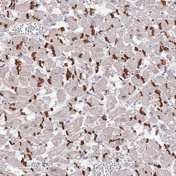
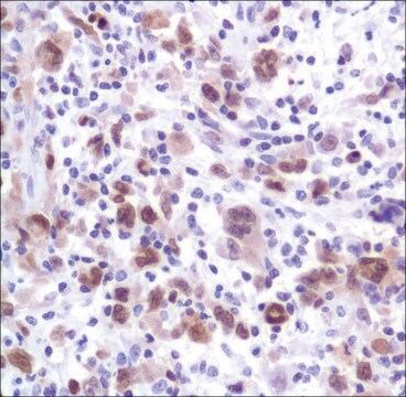
MUM1 (multiple myeloma oncogene-1)/IRF4 (interferon regulatory factor 4) is a 50 kDa protein encoded by MUM1 gene, and a member of the interferon regulatory factor family of transcription factors. MUM1/IRF4 is expressed in the nuclei of plasma cells and a small percentage of germinal center (GC) B-cells located in the “light zone”. This antibody stains MUM1 protein, which is expressed in a subset of B-cells in the light zone of the germinal center, plasma cells, activated T-cells, and a wide spectrum of related hematolymphoid neoplasms derived from these cells. Therefore, this antibody is useful for the subclassiffication of lymphoid malignancies and an excellent marker for Reed-Sternberg cells of classic Hodgkin disease in combination with anti-CD30.

Qualität


IVD

IVD

IVD

RUO

Verlinkung

MUM1 Positive Control Slides, Product No. 358S, are available for immunohistochemistry (formalin-fixed, paraffin-embedded sections).

Physikalische Form

Solution in Tris Buffer, pH 7.3-7.7, with 1% BSA and <0.1% Sodium Azide

Angaben zur Herstellung

Download the IFU specific to your product lot and formatNote: This requires a keycode which can be found on your packaging or product label.

Sonstige Hinweise

For Technical Service please contact: 800-665-7284 or email: service@cellmarque.com

Rechtliche Hinweise

Cell Marque is a trademark of Merck KGaA, Darmstadt, Germany

Sie haben nicht das passende Produkt gefunden?  

Probieren Sie unser Produkt-Auswahlhilfe. aus.


Analysenzertifikate (COA)

Suchen Sie nach Analysenzertifikate (COA), indem Sie die Lot-/Chargennummer des Produkts eingeben. Lot- und Chargennummern sind auf dem Produktetikett hinter den Wörtern ‘Lot’ oder ‘Batch’ (Lot oder Charge) zu finden.

Besitzen Sie dieses Produkt bereits?

In der Dokumentenbibliothek finden Sie die Dokumentation zu den Produkten, die Sie kürzlich erworben haben.

Die Dokumentenbibliothek aufrufen

G Gaidano et al.
Leukemia, 14(4), 563-566 (2000-04-14)
In recent times, the field of B cell lymphoma histogenesis has progressed rapidly due to the increasing availability of histogenetic markers. Genotypic markers of B cell histogenesis are represented by mutations of IgV and BCL-6 genes, which are somatically acquired
Y Natkunam et al.
Modern pathology : an official journal of the United States and Canadian Academy of Pathology, Inc, 14(7), 686-694 (2001-07-17)
The gene encoding MUM1 was characterized as a possible translocation partner in chromosomal abnormalities involving a significant number of multiple myelomas. The overexpression of the MUM1 protein as a result of translocation t(6;14) (p25;q32) identified MUM1 as a putative regulatory
B Falini et al.
Blood, 95(6), 2084-2092 (2000-03-09)
A new monoclonal antibody (MUM1p) was used to study the cell/tissue expression of human MUM1/IRF4 protein, the product of the homologous gene involved in the myeloma-associated t(6;14) (p25;q32). MUM1 was expressed in the nuclei and cytoplasm of plasma cells and
Antonino Carbone et al.
British journal of haematology, 117(2), 366-372 (2002-04-26)
Biological and clinical studies have shown that Hodgkin's disease (HD) can be divided into two major categories, termed nodular lymphocyte predominance HD (NLP HD) and classic HD (CHD). Within CHD four subtypes have been distinguished: nodular sclerosis, mixed cellularity, lymphocyte
Mathewos Tessema et al.
PloS one, 7(4), e34850-e34850 (2012-04-13)
Aberrant cytosine methylation affects regulation of hundreds of genes during cancer development. In this study, a novel aberrantly hypermethylated CpG island in cancer was discovered within the TOX2 promoter. TOX2 was unmethylated in normal cells but 28% lung (n =

Unser Team von Wissenschaftlern verfügt über Erfahrung in allen Forschungsbereichen einschließlich Life Science, Materialwissenschaften, chemischer Synthese, Chromatographie, Analytik und vielen mehr..

Setzen Sie sich mit dem technischen Dienst in Verbindung.