Direkt zum Inhalt
Merck

248M-1

Sigma-Aldrich

Ep-CAM/Epithelial Specific Antigen (MOC-31) Mouse Monoclonal Antibody

Anmeldenzur Ansicht organisationsspezifischer und vertraglich vereinbarter Preise


About This Item

UNSPSC-Code:
12352203
NACRES:
NA.41

Biologische Quelle

mouse

Qualitätsniveau

100
500

Konjugat

unconjugated

Antikörperform

culture supernatant

Antikörper-Produkttyp

primary antibodies

Klon

MOC-31, monoclonal

Beschreibung

For In Vitro Diagnostic Use in Select Regions (See Chart)

Form

buffered aqueous solution

Speziesreaktivität

human

Verpackung

vial of 0.1 mL concentrate (248M-14)
vial of 0.5 mL concentrate (248M-15)
bottle of 1.0 mL predilute (248M-17)
vial of 1.0 mL concentrate (248M-16)
bottle of 7.0 mL predilute (248M-18)

Hersteller/Markenname

Cell Marque

Methode(n)

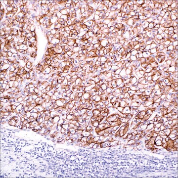
immunohistochemistry (formalin-fixed, paraffin-embedded sections): 1:50-1:200

Isotyp

IgG1κ

Kontrolle

colon carcinoma

Versandbedingung

wet ice

Lagertemp.

2-8°C

Visualisierung

cytoplasmic

Angaben zum Gen

human ... EPCAM(4072)

Allgemeine Beschreibung

Epithel-Zelladhäsionsmolekül (Ep-CAM) ist ein transmembranes Glykoprotein, das auf der Zellmembran der meisten Epithelgewebe lokalisiert ist. Bei den meisten Epithelneoplasien wurde eine Immunreaktivität mit dem Antikörper für Ep-CAM festgestellt. Die meisten Nicht-Epithel-Neoplasien weisen jedoch keine Expression von Ep-CAM auf. Ep-CAM wird in Mesothelzellen, Hepatozyten und Lymphozyten nicht exprimiert. In Verbindung mit anderen Markern kann Ep-CAM als Hilfe zum Bestimmen von Neoplasien mit epithelialem Ursprung eingesetzt werden, beispielsweise um zwischen Adenokarzinom und Mesotheliom der Lunge zu unterscheiden.

Qualität


IVD

IVD

IVD

RUO

Verlinkung

Ep-CAM-positive Kontrollobjektträger, Produkt-Nr. 248S, sind für die Immunhistochemie (formalinfixierte Paraffinschnitte) vorgesehen.

Physikalische Form

Lösung in Tris-Puffer, pH 7,3 - 7,7, mit 1 % BSA und <0,1 % Natriumazid

Angaben zur Herstellung

Laden Sie die für Ihre Produktcharge und Ihr Format geltende Gebrauchsanweisung herunterHinweis: Dies erfordert einen Schlüsselcode, der auf Ihrer Verpackung oder Ihrem Produktetikett angegeben ist.

Sonstige Hinweise

Den Technischen Kundendienst erreichen Sie unter: 800-665-7284 oder per E-Mail: service@cellmarque.com

Rechtliche Hinweise

Cell Marque is a trademark of Merck KGaA, Darmstadt, Germany

Sie haben nicht das passende Produkt gefunden?  

Probieren Sie unser Produkt-Auswahlhilfe. aus.


Hier finden Sie alle aktuellen Versionen:

Analysenzertifikate (COA)

Lot/Batch Number

Die passende Version wird nicht angezeigt?

Wenn Sie eine bestimmte Version benötigen, können Sie anhand der Lot- oder Chargennummer nach einem spezifischen Zertifikat suchen.

Besitzen Sie dieses Produkt bereits?

In der Dokumentenbibliothek finden Sie die Dokumentation zu den Produkten, die Sie kürzlich erworben haben.

Die Dokumentenbibliothek aufrufen

James S Lewis et al.
Modern pathology : an official journal of the United States and Canadian Academy of Pathology, Inc, 18(11), 1471-1481 (2005-06-25)
Sarcomatoid carcinomas are rare malignancies which represent poorly differentiated epithelial tumors that may be difficult to recognize as such. While some cases may have obvious epithelial areas, the sarcomatoid areas are poorly distinguishable from true sarcoma at the light microscopic
Murat Gokden et al.
Diagnostic cytopathology, 33(3), 166-172 (2005-08-04)
Differential diagnosis of primary and metastatic carcinomas of the liver can be problematic and may require immunohistochemical work-up. Recently, new immunohistochemical markers have been introduced with promising results in this area. We studied three of these markers, human hepatocyte antibody
Sanjay Kakar et al.
Archives of pathology & laboratory medicine, 131(11), 1648-1654 (2007-11-06)
Immunohistochemistry plays a crucial role in the diagnosis of hepatocellular carcinoma and in its distinction from other primary and metastatic neoplasms. Because limited tissue is available with fine-needle and core biopsies, appropriate selection of antibodies is imperative. To review the
Jonathan L Hecht et al.
Cancer, 108(1), 56-59 (2005-12-06)
It has been shown previously that detection of the epithelial membrane antigen using the mouse monoclonal antibody MOC-31 has diagnostic utility in the distinction between mesothelioma and metastatic carcinoma in body fluids. The current immunohistochemical study confirmed the effectiveness of
J E King et al.
Histopathology, 48(3), 223-232 (2006-01-25)
Immunohistochemistry is frequently employed to aid the distinction between mesothelioma and pulmonary adenocarcinoma metastatic to the pleura, but there is uncertainty as to which antibodies are most useful. We analysed published data in order to establish sensitivity and specificity of

Verwandter Inhalt

Diagnostic immunohistochemistry overview highlights its importance in cancer diagnosis and detecting infectious diseases in modern clinical pathology.

Unser Team von Wissenschaftlern verfügt über Erfahrung in allen Forschungsbereichen einschließlich Life Science, Materialwissenschaften, chemischer Synthese, Chromatographie, Analytik und vielen mehr..

Setzen Sie sich mit dem technischen Dienst in Verbindung.