Direkt zum Inhalt
Merck
Alle Fotos(1)

Key Documents

NE1022

Sigma-Aldrich

PhosphoDetect Anti-Neurofilament H Mouse mAb (SMI-31)

liquid, clone SMI-31, Calbiochem®

Synonym(e):

Anti-neurofilament antibody

Anmeldenzur Ansicht organisationsspezifischer und vertraglich vereinbarter Preise


About This Item

UNSPSC-Code:
12352203
NACRES:
NA.41

Biologische Quelle

mouse

Qualitätsniveau

Antikörperform

purified antibody

Antikörper-Produkttyp

primary antibodies

Klon

SMI-31, monoclonal

Form

liquid

Enthält

≤0.1% sodium azide as preservative

Speziesreaktivität

chicken, Xenopus

Speziesreaktivität (Voraussage durch Homologie)

mammals

Hersteller/Markenname

Calbiochem®

Lagerbedingungen

OK to freeze
avoid repeated freeze/thaw cycles

Isotyp

IgG1

Versandbedingung

wet ice

Lagertemp.

2-8°C

Posttranslationale Modifikation Target

unmodified

Angaben zum Gen

human ... NEFH(4744)

Allgemeine Beschreibung

Mouse monoclonal antibody supplied as purified antibody. Recognizes the ~180-200 kDa phosphorylated neurofilament H protein.
Recognizes the ~180-200 kDa phosphorylated neurofilament H protein in rat central nervous system (CNS) cytoskeletal preparations. Also weakly recognizes phosphorylated NF-M protein.
This PhosphoDetect Anti-Neurofilament H Mouse mAb (SMI-31) is validated for use in ELISA, Frozen Sections, Immunoblotting, ICC, Paraffin Sections, IP for the detection of Neurofilament H.

Immunogen

homogenized, hypothalami from Fischer 344 rat brain

Anwendung


ELISA (1:1000)
Frozen Sections (1:1000, see comments)
Immunoblotting (1:1000)
Immunocytochemistry (1:1000, see comments)
Paraffin Sections (1:1000, heat pre-treatment required, see comments)
Immunoprecipitation (see comments)

Warnhinweis

Toxicity: Standard Handling (A)

Physikalische Form

In PBS and Thimerosal

Sonstige Hinweise

Raina, A.K., et al. 1999. Neuroreport10, 1355.
Yang, C.C., et al. 1998. Brain121, 1089.
Giasson, B.I and Mushynski, W.E. 1996. J. Biol. Chem.271, 30404.
Mirabella, M., et al. 1996. J. Neuropath. Exp. Neurol.55, 774.
Xiao, J. and Monteiro, M.J. 1994. J. Neurosci.14, 1820.
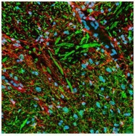
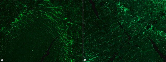
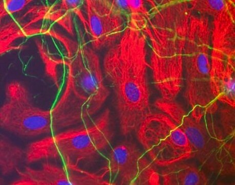
Strongly recognizes phosphorylated neurofilament H and, to a lesser extent, phosphorylated neurofilament M. By immunocytochemistry this antibody broadly stains thick and thin axons and some dendrites, such as basket cell dendrites, but not Purkinje cell dendrites. Does not generally stain nerve cell bodies or other cells and tissues except peripheral axons. May also stain neuronal cell bodies in pathological conditions. Aberrant phosphorylation of neurofilament H in cell bodies can be demonstrated in neuronal cell cultures following treatment with agents that induce stress-activated protein kinase. This antibody is reported to co-immunoprecipitate neurofilament-associated kinase (NAK 115) via interaction of the antibody with the tail domain of neurofilament H. Phosphatase treatment of tissue sections or immunoblotting samples abolishes antibody reactivity. Reactivity is unaffected by trypsin treatment of samples. Tissues and cultured cells can be fixed with a variety of paraformaldehyde- or formaldehyde-containing fixatives, including Bouin′s fixative. Post-fixation in cold methanol or methanol/hydrogen peroxide facilitates access of the antibody to the epitope in frozen sections or thick tissue sections fixed in 4% paraformaldehyde and in cultured cells. For staining formalin-fixed, paraffin sections it is recommended that the de-paraffinized tissue be autoclaved in dH2O for 10 min or boiled in Tris-buffered saline, pH 9.0, in a microwave for 15 min to expose the epitope. Antibody should be titrated for optimal results in individual systems.

Rechtliche Hinweise

CALBIOCHEM is a registered trademark of Merck KGaA, Darmstadt, Germany

Sie haben nicht das passende Produkt gefunden?  

Probieren Sie unser Produkt-Auswahlhilfe. aus.

Lagerklassenschlüssel

12 - Non Combustible Liquids

WGK

nwg

Flammpunkt (°F)

Not applicable

Flammpunkt (°C)

Not applicable


Analysenzertifikate (COA)

Suchen Sie nach Analysenzertifikate (COA), indem Sie die Lot-/Chargennummer des Produkts eingeben. Lot- und Chargennummern sind auf dem Produktetikett hinter den Wörtern ‘Lot’ oder ‘Batch’ (Lot oder Charge) zu finden.

Besitzen Sie dieses Produkt bereits?

In der Dokumentenbibliothek finden Sie die Dokumentation zu den Produkten, die Sie kürzlich erworben haben.

Die Dokumentenbibliothek aufrufen

Masamitsu Shimazawa et al.
Journal of neuroscience research, 90(10), 1960-1969 (2012-06-08)
We evaluated time-dependent optic nerve degeneration and the role of endoplasmic reticulum (ER) stress in this process following retinal ganglion cell death in mice. Retinal damage was induced by intravitreal injection of N-methyl-D-aspartate (NMDA). Neurofilament heavy (NFH)- and phosphorylated NFH
Seungyong Lee et al.
Nature communications, 12(1), 4939-4939 (2021-08-18)
Pain is a central feature of soft tissue trauma, which under certain contexts, results in aberrant osteochondral differentiation of tissue-specific stem cells. Here, the role of sensory nerve fibers in this abnormal cell fate decision is investigated using a severe
Justin C Burrell et al.
Journal of biomedical materials research. Part A, 109(7), 1183-1195 (2020-09-29)
Promising biomaterials should be tested in appropriate large animal models that recapitulate human inflammatory and regenerative responses. Previous studies have shown tyrosine-derived polycarbonates (TyrPC) are versatile biomaterials with a wide range of applications across multiple disciplines. The library of TyrPC
Chongbo Zhong et al.
Frontiers in neural circuits, 16, 978837-978837 (2022-10-11)
Modulation of the release of glutamate by activation of presynaptic nicotinic acetylcholine receptors (nAChRs) is one of the most prevalent mechanism of nicotinic facilitation of glutamatergic transmission in cortico-limbic circuits. By imaging gene chimeric co-cultures from mouse, we examined the
Michael J Whitehead et al.
Scientific reports, 8(1), 5219-5219 (2018-03-28)
Axon degeneration underlies many nervous system diseases; therefore understanding the regulatory signalling pathways is fundamental to identifying potential therapeutics. Previously, we demonstrated heparan sulphates (HS) as a potentially new target for promoting CNS repair. HS modulate cell signalling by both

Unser Team von Wissenschaftlern verfügt über Erfahrung in allen Forschungsbereichen einschließlich Life Science, Materialwissenschaften, chemischer Synthese, Chromatographie, Analytik und vielen mehr..

Setzen Sie sich mit dem technischen Dienst in Verbindung.