Direkt zum Inhalt
Merck
Alle Fotos(1)

Wichtige Dokumente

MABF2778

Sigma-Aldrich

Anti-IRF-3 Antibody, clone AR-1

Anmeldenzur Ansicht organisationsspezifischer und vertraglich vereinbarter Preise


About This Item

UNSPSC-Code:
12352203
NACRES:
NA.41
Klon:
AR-1, monoclonal
application:
ELISA
FACS
IP
WB
inhibition assay
Speziesreaktivität:
rhesus macaque, human
Methode(n):
ELISA: suitable
flow cytometry: suitable
immunoprecipitation (IP): suitable
inhibition assay: suitable
western blot: suitable
citations:

Biologische Quelle

mouse

Qualitätsniveau

Konjugat

unconjugated

Antikörperform

purified antibody

Antikörper-Produkttyp

primary antibodies

Klon

AR-1, monoclonal

Mol-Gew.

calculated mol wt 47.22 kDa
observed mol wt ~51 kDa

Speziesreaktivität

rhesus macaque, human

Verpackung

antibody small pack of 100 μL

Methode(n)

ELISA: suitable
flow cytometry: suitable
immunoprecipitation (IP): suitable
inhibition assay: suitable
western blot: suitable

Isotyp

IgG1

UniProt-Hinterlegungsnummer

Versandbedingung

dry ice

Lagertemp.

2-8°C

Posttranslationale Modifikation Target

unmodified

Allgemeine Beschreibung

Interferon regulatory factor 3 (UniProt: Q14653; also known as IRF-3) is encoded by the IRF3 gene (Gene ID: 3661) in human. IRF-3 is a transcription factor that is constitutively expressed in a variety of tissues and plays a role in intracellular immune response against DNA and RNA viruses. It regulates the transcription of type I interferon genes (IFN-a and IFN-b) and interferon-stimulated genes by binding to an interferon-stimulated response element in their promoters. IRF-3 contains a DNA-binding domain (aa 5-111), nuclear export signal (aa 139-149), IRF-interacting domain, and a C-terminal serine-rich region that contains several phosphorylation sites. Although some of these serines are shown to be phosphorylated in the resting state, further phosphorylation of specific residues is required for its activation and nuclear translocation. In uninfected cells, IRF-3 is present in the cytoplasm, however, following infection, it undergoes phosphorylation on serine 385, serine 386, threonine 390, and serine 396. These phosphorylation events induce a conformational change that leads to its dimerization and nuclear localization where it associates with CREB binding protein to form dsRNA activated factor 1 that subsequently leads to the activation of type I interferons and ISG genes. Phosphorylation and subsequent activation of IRF-3 shown to be inhibited by vaccinia virus protein E3. Mutations in IRF3 gene have been linked to infection-induced, Herpes-specific acute encephalopathy that is characterized by hemorrhagic necrosis of parts of the temporal and frontal lobes. Clone AR-1 recognizes an epitope within the DNA-binding domain of IRF-3. It can detect resting IRF-3 as well as the activated form. (Ref.: Rustagi, A., et al. (2013). Methods. 59(2); 225-232; Honda, K., et al. (2006). Immunity. 25(3); 349-360).

Spezifität

Clone AR1 is a mouse monoclonal antibody that detects Interferon regulatory factor 3 (IRF-3). It targets an epitope within the DNA-binding domain from the N-terminal half.

Immunogen

His-tagged full-length human recombinant human Interferon regulatory factor 3 (IRF-3).

Anwendung

Quality Control Testing

Evaluated by Western Blotting in A549 cell lysate.

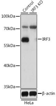
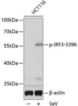
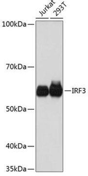
Western Blotting Analysis: A 1:500 dilution of this antibody detected IRF-3 in A549 cell lysate.

Tested Applications

ELISA Analysis: A representative lot detected IRF-3 in ELISA applications (Rustagi, A., et. al. (2013). Methods. 59(2):225-32).

Flow Cytometry Analysis: A representative lot detected IRF-3 in Flow Cytometry applications (Rustagi, A., et. al. (2013). Methods. 59(2):225-32).

Western Blotting Analysis: A representative lot detected IRF-3 in Western Blotting applications (Rustagi, A., et. al. (2013). Methods. 59(2):225-32).

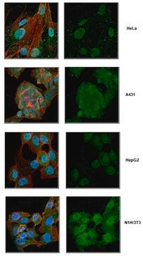
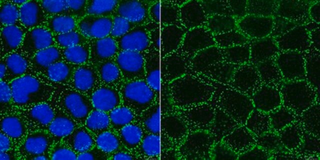
Immunocytochemistry Analysis: A representative lot detected IRF-3 in Immunocytochemistry applications (Rustagi, A., et. al. (2013). Methods. 59(2):225-32).

Note: Actual optimal working dilutions must be determined by end user as specimens, and experimental conditions may vary with the end user
Anti-IRF-3, clone AR-1, Cat. No. MABF2778, is a mouse monoclonal antibody that detects Interferon regulatory factor 3 and is tested for use in ELISA, Flow Cytometry, Immunocytochemistry, and Western Blotting.

Physikalische Form

Purified mouse monoclonal antibody IgG1 in buffer containing 0.1 M Tris-Glycine (pH 7.4), 150 mM NaCl with 0.05% sodium azide.

Lagerung und Haltbarkeit

Recommend storage at +2°C to +8°C. For long term storage antibodies can be kept at -20°C. Avoid repeated freeze-thaws.

Sonstige Hinweise

Concentration: Please refer to the Certificate of Analysis for the lot-specific concentration.

Haftungsausschluss

Unless otherwise stated in our catalog or other company documentation accompanying the product(s), our products are intended for research use only and are not to be used for any other purpose, which includes but is not limited to, unauthorized commercial uses, in vitro diagnostic uses, ex vivo or in vivo therapeutic uses or any type of consumption or application to humans or animals.

Sie haben nicht das passende Produkt gefunden?  

Probieren Sie unser Produkt-Auswahlhilfe. aus.

Lagerklassenschlüssel

12 - Non Combustible Liquids

WGK

WGK 1

Flammpunkt (°F)

Not applicable

Flammpunkt (°C)

Not applicable


Analysenzertifikate (COA)

Suchen Sie nach Analysenzertifikate (COA), indem Sie die Lot-/Chargennummer des Produkts eingeben. Lot- und Chargennummern sind auf dem Produktetikett hinter den Wörtern ‘Lot’ oder ‘Batch’ (Lot oder Charge) zu finden.

Besitzen Sie dieses Produkt bereits?

In der Dokumentenbibliothek finden Sie die Dokumentation zu den Produkten, die Sie kürzlich erworben haben.

Die Dokumentenbibliothek aufrufen

Unser Team von Wissenschaftlern verfügt über Erfahrung in allen Forschungsbereichen einschließlich Life Science, Materialwissenschaften, chemischer Synthese, Chromatographie, Analytik und vielen mehr..

Setzen Sie sich mit dem technischen Dienst in Verbindung.