Spezifität
Clone 3F3-FMA is a mouse monoclonal antibody that detects human Transferrin receptor protein 1.
Immunogen
Purified membrane fractions from OCI-LY7 cells.
Anwendung
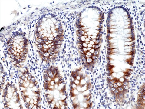
Quality Control TestingEvaluated by Western Blotting in HT-1080 cell lysate.Western Blotting Analysis: A 1:500 dilution of this antibody detected TfR1 (CD71) in HT-1080 cell lysate.Tested ApplicationsFlow Cytometry Analysis: A representative lot detected TfR1 (CD71) in Flow Cytometry applications (Feng, H., et. al. (2020). Cell Rep. 30(10): 3411-3423).Immunocytochemistry Analysis: A 1:1,000 dilution from a representative lot detected TfR1 (CD71) in HT-1080 cells.Immunohistochemistry Applications: A representative lot detected TfR1 (CD71) in Immunohistochemistry applications (Feng, H., et. al. (2020). Cell Rep. 30(10): 3411-3423).Immunocytochemistry Analysis: A representative lot detected TfR1 (CD71) in Immunocytochemistry applications (Feng, H., et. al. (2020). Cell Rep. 30(10): 3411-3423). Western Blotting Analysis: A representative lot detected TfR1 (CD71) in Western Blotting applications (Feng, H., et. al. (2020). Cell Rep. 30(10): 3411-3423). Note: Actual optimal working dilutions must be determined by end user as specimens, and experimental conditions may vary with the end user
Anti-TfR1 (CD71), clone 3F3-FMA, Cat. No. MABC1765, is a mouse monoclonal antibody that detects Tfr1 and is an excellent ferroptosis marker. It is tested for use in Flow Cytometry, Immunocytochemistry, Immunohistochemistry, and Western Blotting.
Zielbeschreibung
Transferrin receptor protein 1 (UniProt: Q62351; also known as TR, TfR, TfR1, Trfr, T9, p90, CD71) is encoded by the Tfrc (also known as Trfr) gene (Gene ID: 7037) in human. TfR1 is a single-pass type II membrane glycoprotein with a cytoplasmic domain (aa 1-67), a transmembrane domain (aa 68-88), and a long extracellular domain (aa 89-760). It can also be proteolytically cleaved at Arg100 to generate the soluble serum form (sTfR). TfR1 is involved in the uptake of iron via receptor mediated endocytosis into specialized endosomes where acidification leads to release of iron. TfR1 is reported to be essential for erythrocyte development and positive regulation of T and B cell proliferation through iron uptake. It also acts as a lipid sensor that regulates mitochondrial fusion by regulating the activation of the JNK pathway and degradation of mitofusin MFN2. Under conditions of low dietary levels of stearate (C18:0), it promotes the activation of JNK pathway that results in HUWE1-mediated ubiquitination and degradation of mitofusin MFN2 and inhibition of mitochondrial fusion. Clone 3F3-FMA is reported to be an effective ferroptosis-staining agent. It stains plasma membrane and the perinuclear region associated with the Golgi and the endosomal recycling compartment. (Ref.: Senilymaz, D., et al. (2015). Nature. 525(7567); 124-128; Feng, H., et al. (2020). Cell Rep. 30(10); 3411-3423).
Physikalische Form
Purified mouse monoclonal antibody IgG1 in buffer containing 0.1 M Tris-Glycine (pH 7.4), 150 mM NaCl with 0.05% sodium azide.
Lagerung und Haltbarkeit
Stable for 1 year at +2°C to +8°C from date of receipt.
Sonstige Hinweise
Concentration: Please refer to the Certificate of Analysis for the lot-specific concentration.
Haftungsausschluss
Unless otherwise stated in our catalog or other company documentation accompanying the product(s), our products are intended for research use only and are not to be used for any other purpose, which includes but is not limited to, unauthorized commercial uses, in vitro diagnostic uses, ex vivo or in vivo therapeutic uses or any type of consumption or application to humans or animals.
