Skip to Content
Merck
All Photos(6)

Key Documents

SAB2700790

Sigma-Aldrich

Anti-SLC32A1 (N-terminal) antibody produced in rabbit

affinity isolated antibody, buffered aqueous solution

Synonym(s):

SLC32A1, VGAT, VIAAT

Sign Into View Organizational & Contract Pricing


About This Item

UNSPSC Code:
12352203
NACRES:
NA.41

biological source

rabbit

Quality Level

conjugate

unconjugated

antibody form

affinity isolated antibody

antibody product type

primary antibodies

clone

polyclonal

form

buffered aqueous solution

species reactivity

mouse, rat, human

technique(s)

immunofluorescence: suitable
immunohistochemistry: suitable
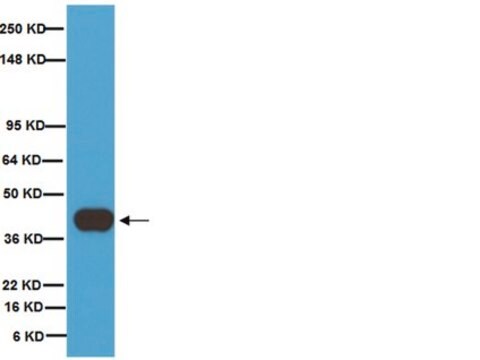
western blot: 1000-10000

NCBI accession no.

UniProt accession no.

shipped in

wet ice

storage temp.

−20°C

Gene Information

Immunogen

Recombinant fragment corresponding to a region within amino acids 1 and 118 of VGAT according to NP_542119

Application

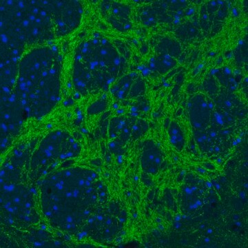
Suggested starting dilutions are as follows: ICC/IF: 1:100-1:1000, IHC-Fr: 1:100-1:1000, IHC-P: 1:100-1:1000, WB: 1:1000-1:10000. Not yet tested in other applications. Optimal working dilutions should be determined experimentally by the end user.

Biochem/physiol Actions

The protein encoded by this gene is an integral membrane protein involved in gamma-aminobutyric acid (GABA) and glycine uptake into synaptic vesicles. The encoded protein is a member of amino acid/polyamine transporter family II. [provided by RefSeq]

Features and Benefits

Evaluate our antibodies with complete peace of mind. If the antibody does not perform in your application, we will issue a full credit or replacement antibody. Learn more.

Physical form

1XPBS, 1% BSA, 20% Glycerol (pH7). 0.01% Thimerosal was added as a preservative.

Disclaimer

Unless otherwise stated in our catalog or other company documentation accompanying the product(s), our products are intended for research use only and are not to be used for any other purpose, which includes but is not limited to, unauthorized commercial uses, in vitro diagnostic uses, ex vivo or in vivo therapeutic uses or any type of consumption or application to humans or animals.

Not finding the right product?  

Try our Product Selector Tool.

Pictograms

Exclamation mark

Signal Word

Warning

Hazard Statements

Hazard Classifications

Aquatic Chronic 3 - Skin Sens. 1

Storage Class Code

12 - Non Combustible Liquids

WGK

WGK 2

Flash Point(F)

Not applicable

Flash Point(C)

Not applicable


Certificates of Analysis (COA)

Search for Certificates of Analysis (COA) by entering the products Lot/Batch Number. Lot and Batch Numbers can be found on a product’s label following the words ‘Lot’ or ‘Batch’.

Already Own This Product?

Find documentation for the products that you have recently purchased in the Document Library.

Visit the Document Library

Borislav Dejanovic et al.
Neurobiology of disease, 67, 88-96 (2014-02-25)
Gephyrin is a postsynaptic scaffolding protein, essential for the clustering of glycine and γ-aminobutyric acid type-A receptors (GABAARs) at inhibitory synapses. An impairment of GABAergic synaptic inhibition represents a key pathway of epileptogenesis. Recently, exonic microdeletions in the gephyrin (GPHN)
Joshua J White et al.
The Journal of neuroscience : the official journal of the Society for Neuroscience, 34(24), 8231-8245 (2014-06-13)
Cerebellar circuits are patterned into an array of topographic parasagittal domains called zones. The proper connectivity of zones is critical for motor coordination and motor learning, and in several neurological diseases cerebellar circuits degenerate in zonal patterns. Despite recent advances
Shih-Hsiu J Wang et al.
The Journal of neuroscience : the official journal of the Society for Neuroscience, 34(38), 12745-12761 (2014-09-19)
Most excitatory synapses in the mammalian brain are formed on dendritic spines, and spine density has a profound impact on synaptic transmission, integration, and plasticity. Membrane-associated guanylate kinase (MAGUK) proteins are intracellular scaffolding proteins with well established roles in synapse
Ali Mehanna et al.
Experimental neurology, 261, 147-155 (2014-06-27)
The immune system plays important functional roles in regeneration after injury to the mammalian central and peripheral nervous systems. After damage to the peripheral nerve several types of immune cells, invade the nerve within hours after the injury. To gain
Zoé Husson et al.
The Journal of neuroscience : the official journal of the Society for Neuroscience, 34(28), 9418-9431 (2014-07-11)
The principal neurons of the cerebellar nuclei (CN), the sole output of the olivo-cerebellar system, receive a massive inhibitory input from Purkinje cells (PCs) of the cerebellar cortex. Morphological evidence suggests that CN principal cells are also contacted by inhibitory

Our team of scientists has experience in all areas of research including Life Science, Material Science, Chemical Synthesis, Chromatography, Analytical and many others.

Contact Technical Service