Skip to Content
Merck
All Photos(1)

Key Documents

P4850

Sigma-Aldrich

Proteinase K from Tritirachium album

buffered aqueous glycerol solution, for molecular biology, ≥800 units/mL

Synonym(s):

Endopeptidase K

Sign Into View Organizational & Contract Pricing


About This Item

CAS Number:
Enzyme Commission number:
MDL number:
UNSPSC Code:
12352204
NACRES:
NA.54

grade

for molecular biology

Quality Level

form

buffered aqueous glycerol solution

mol wt

28.93 kDa

concentration

≥10 mg/mL
≥800 units/mL

impurities

≤0.5 ppm DNA (PicoGreen® assay)

foreign activity

DNase, Nickase and RNase, none detected

storage temp.

2-8°C

Looking for similar products? Visit Product Comparison Guide

Application

Proteinase K from Tritirachium album has been used:
  • in bovine endometrial epithelial cells for herpes viral DNA extraction
  • for viral RNA extraction from nasal swabs
  • as a component of phase lock and direct PCR lysis buffer

The product has been used to study its pre-treatment effects on the silk fibroin. The aspects analysed in this study included the crystallographic properties of hydroxyapatite (HAp), and the microstructure and microhardness of the composites. The enzyme has also been used to facilitate the access of probes to rRNA using FISH techniques to detect pathogenic Staphylococcus aureus.
Useful for the proteolytic inactivation of nucleases during the isolation of DNA and RNA.
Removes endotoxins that bind to cationic proteins such as lysozyme and ribonuclease A.
Reported useful for the isolation of hepatic, yeast, and mung bean mitochondria
Determination of enzyme localization on membranes
Treatment of paraffin embedded tissue sections to expose antigen binding sites for antibody labeling.
Digestion of proteins from brain tissue samples for prions in Transmissible Spongiform Encephalopathies (TSE) research.

Biochem/physiol Actions

Proteinase K has a broad specificity and degrades many proteins even in the native state. It mainly cleaves the peptide bond adjacent to the carboxyl group of aliphatic and aromatic amino acids with blocked α-amino groups. The molecular weight of proteinase K from amino acid sequence is found to be 28,930 Da and from SDS-PAGE, it is found to be 28,500 Da. The optimum pH is between 7.5-9.0 and its isoelectric point is 8.9. Ca2+ (1-5 mM) is required for its activation. Proteinase K is inhibited by DIFP (diisopropylfluorophosphate) or PMSF (phenylmethylsulfonyl fluoride).
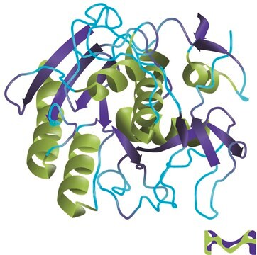
Proteinase K is a stable and highly reactive serine protease. Evidence from crystal and molecular structure studies indicates the enzyme belongs to the subtilisin family with an active-site catalytic triad (Asp39-His69-Ser224). It is stable in a broad range of environments: pH, buffer salts, detergents (SDS), and temperature. In the presence of 0.1-0.5% SDS, proteinase K retains activity and will digest a variety of proteins and nucleases in DNA preparations without compromising the integrity of the isolated DNA.

Unit Definition

One unit will hydrolyze urea-denatured hemoglobin to produce color equivalent to 1.0 μmole of tyrosine per min at pH 7.5 at 37 °C (color by Folin-Ciocalteu reagent).

Physical form

Solution in 40% glycerol (v/v) containing 10 mM Tris-HCl, pH 7.5, with 1 mM calcium acetate.

Legal Information

PicoGreen is a registered trademark of Life Technologies

Pictograms

Health hazard

Signal Word

Danger

Hazard Statements

Precautionary Statements

Hazard Classifications

Resp. Sens. 1

Storage Class Code

10 - Combustible liquids

WGK

WGK 2

Flash Point(F)

Not applicable

Flash Point(C)

Not applicable

Personal Protective Equipment

dust mask type N95 (US), Eyeshields, Gloves

Certificates of Analysis (COA)

Search for Certificates of Analysis (COA) by entering the products Lot/Batch Number. Lot and Batch Numbers can be found on a product’s label following the words ‘Lot’ or ‘Batch’.

Already Own This Product?

Find documentation for the products that you have recently purchased in the Document Library.

Visit the Document Library

Bovine herpes virus type 4 alters TNF-alpha and IL-8 profiles and impairs the survival of bovine endometrial epithelial cells
Chanrot M, et al.
Reproductive Biology, 17(3), 225-232 (2017)
Shin-Rong J Wu et al.
Scientific reports, 9(1), 11434-11434 (2019-08-09)
The highly conserved SNARE protein SEC22B mediates diverse and critical functions, including phagocytosis, cell growth, autophagy, and protein secretion. However, these characterizations have thus far been limited to in vitro work. Here, we expand our understanding of the role Sec22b
Robust Generation of Knock-in Cell Lines Using CRISPR-Cas9 and rAAV-assisted Repair Template Delivery
Vandemoortele G, et al.
Bio-protocol, 7(7), 1-6 (2017)
A S Neimanis et al.
Transboundary and emerging diseases, 65(1), 213-220 (2017-04-14)
Incursion of rabbit haemorrhagic disease virus (RHDV) into Sweden was documented in 1990 and it is now considered endemic in wild rabbit (Oryctolagus cuniculus) populations. Rabbit haemorrhagic disease virus 2 (RHDV2), a new, related lagovirus was first detected in France
Lorrayne Serra et al.
G3 (Bethesda, Md.), 9(8), 2687-2697 (2019-05-23)
Entomopathogenic nematodes from the genus Steinernema are lethal insect parasites that quickly kill their insect hosts with the help of their symbiotic bacteria. Steinernema carpocapsae is one of the most studied entomopathogens due to its broad lethality to diverse insect

Articles

Proteinase K (EC 3.4.21.64) activity can be measured spectrophotometrically using hemoglobin as the substrate. Proteinase K hydrolyzes hemoglobin denatured with urea, and liberates Folin-postive amino acids and peptides. One unit will hydrolyze hemoglobin to produce color equivalent to 1.0 μmol of tyrosine per minute at pH 7.5 at 37 °C (color by Folin & Ciocalteu's Phenol Reagent).

Protocols

Proteinase K (EC 3.4.21.64) activity can be measured spectrophotometrically using hemoglobin as the substrate. Proteinase K hydrolyzes hemoglobin denatured with urea, and liberates Folin-postive amino acids and peptides. One unit will hydrolyze hemoglobin to produce color equivalent to 1.0 μmol of tyrosine per minute at pH 7.5 at 37 °C (color by Folin & Ciocalteu's Phenol Reagent).

Our team of scientists has experience in all areas of research including Life Science, Material Science, Chemical Synthesis, Chromatography, Analytical and many others.

Contact Technical Service