Skip to Content
Merck
All Photos(13)

Key Documents

HPA014341

Sigma-Aldrich

Anti-MS4A1 antibody produced in rabbit

enhanced validation

Prestige Antibodies® Powered by Atlas Antibodies, affinity isolated antibody, buffered aqueous glycerol solution

Synonym(s):

Anti-B-lymphocyte antigen CD20, Anti-B-lymphocyte surface antigen B1, Anti-Bp35, Anti-Leu-16, Anti-Membrane-spanning 4-domains subfamily A member 1

Sign Into View Organizational & Contract Pricing


About This Item

UNSPSC Code:
12352203
Human Protein Atlas Number:
NACRES:
NA.41

biological source

rabbit

Quality Level

conjugate

unconjugated

antibody form

affinity isolated antibody

antibody product type

primary antibodies

clone

polyclonal

product line

Prestige Antibodies® Powered by Atlas Antibodies

form

buffered aqueous glycerol solution

species reactivity

human

enhanced validation

independent
orthogonal RNAseq
Learn more about Antibody Enhanced Validation

technique(s)

immunoblotting: 0.04-0.4 μg/mL
immunofluorescence: 0.25-2 μg/mL
immunohistochemistry: 1:1000-1:2500

immunogen sequence

ENEWKRTCSRPKSNIVLLSAEEKKEQTIEIKEEVVGLTETSSQPKNEEDIEIIPIQEEEEEETETNFPEPPQDQESSP

UniProt accession no.

shipped in

wet ice

storage temp.

−20°C

target post-translational modification

unmodified

Gene Information

human ... MS4A1(931)

General description

The gene MS4A1 (membrane spanning 4-domains A1) is also referred to as CD20 and is expressed on the surface of human B lymphocytes and to some extent on a small subset of T cells. The 33 to 35kDa, nonglycosylated protein has 4 membrane-spanning domains with both the amino and carboxy termini of the protein localized to the cytoplasm. The gene is mapped to human chromosome 11q12-13.

Immunogen

B-lymphocyte antigen CD20 recombinant protein epitope signature tag (PrEST)

Application

All Prestige Antibodies Powered by Atlas Antibodies are developed and validated by the Human Protein Atlas (HPA) project and as a result, are supported by the most extensive characterization in the industry.

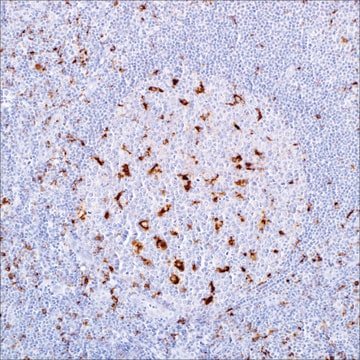
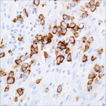
The Human Protein Atlas project can be subdivided into three efforts: Human Tissue Atlas, Cancer Atlas, and Human Cell Atlas. The antibodies that have been generated in support of the Tissue and Cancer Atlas projects have been tested by immunohistochemistry against hundreds of normal and disease tissues and through the recent efforts of the Human Cell Atlas project, many have been characterized by immunofluorescence to map the human proteome not only at the tissue level but now at the subcellular level. These images and the collection of this vast data set can be viewed on the Human Protein Atlas (HPA) site by clicking on the Image Gallery link. We also provide Prestige Antibodies® protocols and other useful information.

Biochem/physiol Actions

The gene MS4A1 (membrane spanning 4-domains A1) encodes a protein belonging to the membrane-spanning 4A gene family that participates in the development and cell cycle progression in B-cells. It has been found to interact with proteins, such as transmembrane adapter protein p75/80, CD40, and major histocompatibility complex class II proteins (MHC II). It may participate in Ca2+ influx across plasma membranes, maintenance of Ca2+ concentration, and activation of B-cells. The MS4A1 protein activates Src leading to rapid phosphorylation of phospholipase C-γ1 (PLC-γ1) and PLC-γ2, in turn activating caspase-3 signaling of apoptosis.

Features and Benefits

Prestige Antibodies® are highly characterized and extensively validated antibodies with the added benefit of all available characterization data for each target being accessible via the Human Protein Atlas portal linked just below the product name at the top of this page. The uniqueness and low cross-reactivity of the Prestige Antibodies® to other proteins are due to a thorough selection of antigen regions, affinity purification, and stringent selection. Prestige antigen controls are available for every corresponding Prestige Antibody and can be found in the linkage section.

Every Prestige Antibody is tested in the following ways:
  • IHC tissue array of 44 normal human tissues and 20 of the most common cancer type tissues.
  • Protein array of 364 human recombinant protein fragments.

Linkage

Corresponding Antigen APREST73138

Physical form

Solution in phosphate-buffered saline, pH 7.2, containing 40% glycerol and 0.02% sodium azide

Legal Information

Prestige Antibodies is a registered trademark of Merck KGaA, Darmstadt, Germany

Disclaimer

Unless otherwise stated in our catalog or other company documentation accompanying the product(s), our products are intended for research use only and are not to be used for any other purpose, which includes but is not limited to, unauthorized commercial uses, in vitro diagnostic uses, ex vivo or in vivo therapeutic uses or any type of consumption or application to humans or animals.

Not finding the right product?  

Try our Product Selector Tool.

Storage Class Code

10 - Combustible liquids

WGK

WGK 1

Flash Point(F)

Not applicable

Flash Point(C)

Not applicable

Personal Protective Equipment

dust mask type N95 (US), Eyeshields, Gloves

Certificates of Analysis (COA)

Search for Certificates of Analysis (COA) by entering the products Lot/Batch Number. Lot and Batch Numbers can be found on a product’s label following the words ‘Lot’ or ‘Batch’.

Already Own This Product?

Find documentation for the products that you have recently purchased in the Document Library.

Visit the Document Library

Ye-Lin Liang et al.
Nature communications, 13(1), 2996-2996 (2022-06-01)
Increasing evidence has revealed the roles of long noncoding RNAs (lncRNAs) as tumor biomarkers. Here, we introduce an immune-associated nine-lncRNA signature for predicting distant metastasis in locoregionally advanced nasopharyngeal carcinoma (LA-NPC). The nine lncRNAs are identified through microarray profiling, followed
Guillaume Cartron et al.
Blood, 104(9), 2635-2642 (2004-07-01)
Rituximab (MabThera, Rituxan) is a chimeric IgG1 monoclonal antibody that specifically targets the CD20 surface antigen expressed on normal and neoplastic B-lymphoid cells. Rituximab is currently used in the treatment of both follicular and aggressive B-cell non-Hodgkin lymphomas. Despite its
Olga Ciccarelli et al.
The Lancet. Neurology, 13(8), 807-822 (2014-07-11)
The mechanisms underlying the pathogenesis of multiple sclerosis induce the changes that underpin relapse-associated and progressive disability. Disease mechanisms can be investigated in preclinical models and patients with multiple sclerosis by molecular and metabolic imaging techniques. Many insights have been
O Aubert et al.
American journal of transplantation : official journal of the American Society of Transplantation and the American Society of Transplant Surgeons, 14(6), 1439-1445 (2014-05-09)
Anti-HLA donor-specific antibodies (DSAs) cause acute and chronic antibody-mediated rejection (AMR). However, the clinical relevance of anti-HLA-C antibodies remains unclear. We evaluated the clinical relevance of the presence of anti-HLA-C DSA at day 0 in renal transplant recipients. In this
Lisa G Rider et al.
Clinical and experimental rheumatology, 32(5), 689-696 (2014-07-30)
We aimed to assess changes in myositis core set measures and ancillary clinical and laboratory data from the National Institutes of Health's subset of patients enrolled in the Rituximab in Myositis trial. Eighteen patients (5 dermatomyositis, 8 polymyositis, 5 juvenile

Our team of scientists has experience in all areas of research including Life Science, Material Science, Chemical Synthesis, Chromatography, Analytical and many others.

Contact Technical Service