Skip to Content
Merck
All Photos(2)

Key Documents

F0162

Sigma-Aldrich

Fibronectin Proteolytic Fragment from human plasma

lyophilized powder, 45 kDa

Synonym(s):

Fibronectin

Sign Into View Organizational & Contract Pricing


About This Item

MDL number:
UNSPSC Code:
12352202
eCl@ss:
42020141
NACRES:
NA.75

biological source

human plasma

Quality Level

Assay

≥90% (SDS-PAGE)

form

lyophilized powder

mol wt

45 kDa

packaging

pkg of 0.5 mg

technique(s)

cell culture | mammalian: suitable

impurities

HIV and HBsAg, source material tested negative
Small proteolytic fragments, may contain traces

solubility

water: soluble ≥0.500 mg/mL, clear to slightly hazy, colorless

UniProt accession no.

shipped in

wet ice

storage temp.

−20°C

Gene Information

human ... FN1(2335)

Looking for similar products? Visit Product Comparison Guide

General description

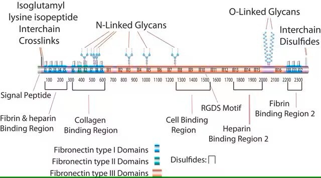
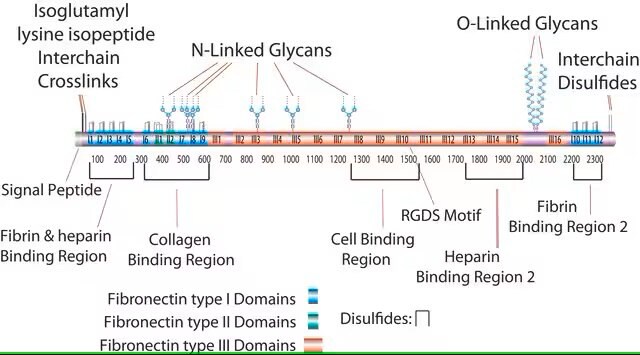
Fibronectin 1 is a glycoprotein of the extracellular matrix that is coded by FN1 gene. It is expressed in the plasma and at the cell surface.FN1 is mapped to human chromosome 2q35. Proteolytic fragments of fibronectin has a cell binding domain. Plasma fibronectin has two polypeptide chains connected by two disulphide bonds present near carboxy terminal. Each polypeptide chain is of 220-250 kDa.

Application

Fibronectin proteolytic fragment from human plasma has been used as fluorophore-conjugated proteins. It has also been used in solid phase binding assay.

Biochem/physiol Actions

Fibronectin participates in cell adhesion, growth, migration, wound healing, blood coagulation and metastasis. Mutations in FN1 results in glomerulopathy. It plays an important role in cell attachment and spreading, control of cell cytoskeleton, morphology and differentiation. FN1 is also involved in extracellular matrix formation, hemostasis and thrombosis. Proteolytic fragments of fibronectin plays a vital role in mononuclear phagocyte function.
Fibronectins are high molecular weight glycoproteins with two subunits joined by a disulfide bond to form the dimer. The fragments are obtained using protelytic enzymes. This 45 kDa gelatin binding fragment is obtained through trypitc digestion of the N-terminal 70 kDa fragment, which is produced by Cathespin D digestion.

This fragment has an acidic pI (4.9-5.3) and does not bind to heparin. This domain is resistant to proteolysis due to intrachain disulfide bonding and the attached carbohydrate. The intrachain disulfide bonds are essential for binding to gelatin, while the complex, branched, asparagine-linked carbohydrate is not. This fragment binds to C1q, but not to fibrin.

Caution

The product should be stored at -20°C.

Preparation Note

This product is lyophilized from phosphate buffered saline with sucrose as a cryoprotectant. The source material has tested negative for antibody to HIV, HCN, and HBsAg. It is soluble in water at 0.5 mg/mL and yields a clear to slightly hazy solution.

Pictograms

Exclamation mark

Signal Word

Warning

Hazard Statements

Hazard Classifications

Eye Irrit. 2 - Skin Irrit. 2 - STOT SE 3

Target Organs

Respiratory system

Storage Class Code

11 - Combustible Solids

WGK

WGK 3

Flash Point(F)

Not applicable

Flash Point(C)

Not applicable


Choose from one of the most recent versions:

Certificates of Analysis (COA)

Lot/Batch Number

Don't see the Right Version?

If you require a particular version, you can look up a specific certificate by the Lot or Batch number.

Already Own This Product?

Find documentation for the products that you have recently purchased in the Document Library.

Visit the Document Library

Nanoparticle amplification via photothermal unveiling of cryptic collagen binding sites.
Lo J H, et al.
Journal of Material Chemistry B: Materials for Biology and Medicine, 1(39), 5235-5240 (2013)
Justin H Lo et al.
Journal of materials chemistry. B, 1(39), 5235-5240 (2013-11-02)
The success of nanoparticle-based cancer therapies ultimately depends on their ability to selectively and efficiently accumulate in regions of disease. Outfitting nanoparticles to actively target tumor-specific markers has improved specificity, yet it remains a challenge to amass adequate therapy in
Fibronectin in Health and Disease (2018)
A R Pickford et al.
Structure (London, England : 1993), 5(3), 359-370 (1997-03-15)
Fibronectin is an extracellular matrix glycoprotein involved in cell adhesion and migration events in a range of important physiological processes. Aberrant adhesion of cells to the matrix may contribute to the breakdown of normal tissue function associated with various diseases.
Plasma Fibronectin: Structure and Functions (1985)

Articles

Extracellular matrix proteins such as laminin, collagen, and fibronectin can be used as cell attachment substrates in cell culture.

Protocols

Dilute fibronectin to the desired concentration. Optimum conditions for attachment are dependent on cell type and application. The typical coating concentration is 1 – 5 ug/cm2.Fibronectin coating protocol, products, and FAQs at sigmaaldrich.com

Dilute fibronectin to the desired concentration. Optimum conditions for attachment are dependent on cell type and application. The typical coating concentration is 1 – 5 ug/cm2.Fibronectin coating protocol, products, and FAQs.

Our team of scientists has experience in all areas of research including Life Science, Material Science, Chemical Synthesis, Chromatography, Analytical and many others.

Contact Technical Service