Skip to Content
Merck
All Photos(1)

Key Documents

C5301

Sigma-Aldrich

Monoclonal Anti-Cytokeratin Peptide 8 antibody produced in mouse

clone M20, ascites fluid

Synonym(s):

Anti-CARD2, Anti-CK-8, Anti-CK8, Anti-CYK8, Anti-K2C8, Anti-K8, Anti-KO

Sign Into View Organizational & Contract Pricing


About This Item

MDL number:
UNSPSC Code:
12352203
NACRES:
NA.41

biological source

mouse

Quality Level

conjugate

unconjugated

antibody form

ascites fluid

antibody product type

primary antibodies

clone

M20, monoclonal

mol wt

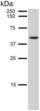
antigen 52 kDa

contains

15 mM sodium azide

species reactivity

feline, human, rabbit, canine, bovine

technique(s)

immunohistochemistry (frozen sections): suitable
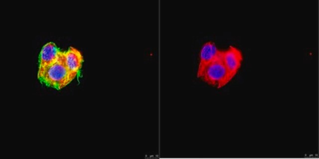
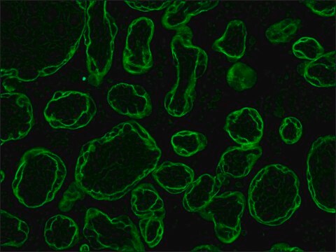
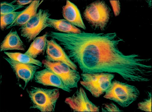
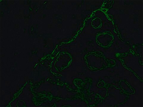
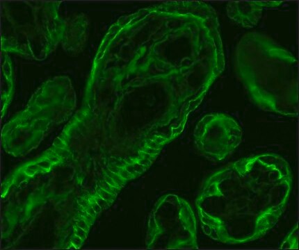
indirect immunofluorescence: 1:200 using frozen sections of human or animal tissue
western blot: suitable

isotype

IgG1

UniProt accession no.

shipped in

dry ice

storage temp.

−20°C

target post-translational modification

unmodified

Gene Information

human ... KRT8(3856)

General description

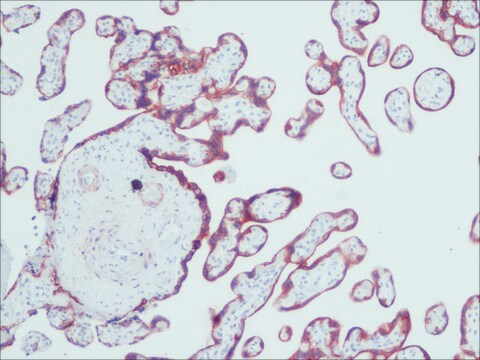
Cytokeratin is a 52.5kD cytoplasmic structural protein that belongs to type II, neutral-to-basic subfamily. It consists of 29 different proteins which is the characteristic of epithelial and trichocytic cells. Monoclonal anti-cytokeratin peptide 8 antibody can be used as a marker for epithelial cell differentiation as well as a tool for tumor identification and classification. It can also be used in western blotting. Mouse anti-cytokeratin peptide 8 antibody reacts specifically with various human simple and complex epithelia like- liver, intestine, pancreas, urinary bladder, salivary gland, thyroid, prostate, mesothel, and placenta but not with stratified squamous epithelia. The product also does not react with non-epithelial tissues (exceptions-certain smooth muscle cells). This product has also shown cross reactivity for cytokeratin of rabbit, cow, dog, and cat.Monoclonal Anti-Cytokeratin 8 (mouse IgG1 isotype) is derived from the M20 hybridoma produced by the fusion of mouse myeloma cells and splenocytes from BALB/c mice immunized with a cytokeratin preparation purified from the human breast carcinoma cell line MCF7.

Specificity

Mouse anti-cytokeratin peptide 8 antibody reacts specifically with various human simple and complex epithelia like- liver, intestine, pancreas, urinary bladder, salivary gland, thyroid, prostate, mesothel, and placenta but not with stratified squamous epithelia. The product also does not react with non-epithelial tissues (exceptions-certain smooth muscle cells). This product has also shown cross reactivity for cytokeratin of rabbit, cow, dog, and cat.

Immunogen

cytokeratin from human breast carcinoma cell line MCF7.

Application

Monoclonal anti-cytokeratin peptide 8 antibody has been used in
  • immunofluorescence microscopy
  • immunohistochemistry
  • dot blotting
  • protein preparation from bovine epithelial cells
  • immunofluorescence
  • western blotting

Biochem/physiol Actions

Monoclonal anti-cytokeratin peptide 8 antibody can be used as a marker for epithelial cell differentiation as well as a tool for tumor identification and classification. It can also be used in western blotting.

Disclaimer

Unless otherwise stated in our catalog or other company documentation accompanying the product(s), our products are intended for research use only and are not to be used for any other purpose, which includes but is not limited to, unauthorized commercial uses, in vitro diagnostic uses, ex vivo or in vivo therapeutic uses or any type of consumption or application to humans or animals.

Not finding the right product?  

Try our Product Selector Tool.

Storage Class Code

10 - Combustible liquids

Flash Point(F)

Not applicable

Flash Point(C)

Not applicable


Certificates of Analysis (COA)

Search for Certificates of Analysis (COA) by entering the products Lot/Batch Number. Lot and Batch Numbers can be found on a product’s label following the words ‘Lot’ or ‘Batch’.

Already Own This Product?

Find documentation for the products that you have recently purchased in the Document Library.

Visit the Document Library

Myoglobin: a promising exogenous reference marker using in proteomics analysis
Wang T, et al.
Food Science and Biotechnology, 22, 393-398 (2013)
David Soler et al.
Genes, chromosomes & cancer, 44(4), 339-350 (2005-07-30)
The development of genomic instability is an important step toward generating the multiple genetic changes required for cancer. Telomere dysfunction is one of the factors that contribute to tumorigenesis. Telomeres shorten with each cell division in the absence of telomerase.
G N Van Muijen et al.
Laboratory investigation; a journal of technical methods and pathology, 57(4), 359-369 (1987-10-01)
Tissues from human fetuses (12 to 14 weeks) were studied by using immunohistochemical methods, with special emphasis on coexpression of intermediate filaments. Well-characterized antibodies, monoclonal as well as polyclonal were used. Indirect immunoperoxidase staining disclosed simultaneous expression of cytokeratin and
S Portet et al.
Cytometry, 35(3), 203-213 (1999-03-19)
In the MCF7 human breast cancer cell line, several patterns of cytokeratin networks are observed, depending on the intracellular localization. Our hypothesis is that architectural variations of cytokeratin networks depend on local tensions or forces appearing spontaneously in the cytoplasm.
Mingxi Hua et al.
Journal of cell science, 125(Pt 23), 5800-5810 (2012-09-15)
The correct functioning of hepatocytes requires the establishment and maintenance of hepatocyte polarity. However, the mechanisms regulating the generation of hepatocyte polarity are not completely understood. The differentiation of human fetal hepatic progenitor cells (hFHPCs) into functional hepatocytes provides a

Our team of scientists has experience in all areas of research including Life Science, Material Science, Chemical Synthesis, Chromatography, Analytical and many others.

Contact Technical Service