306-05F
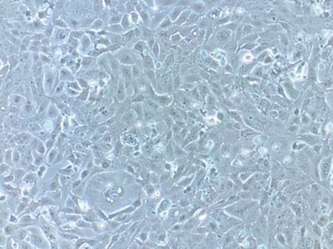
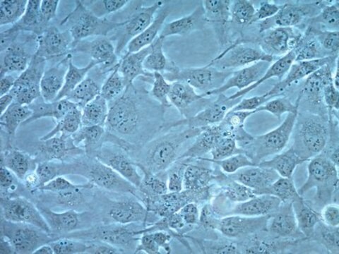
Human Cardiac Fibroblasts: HCF, fetal
Synonym(s):
Cardiac Fibroblasts, Fetal Cardiac Fibroblasts, HCF Cardiac Fibroblasts
About This Item
Recommended Products
biological source
human heart (normal)
Quality Level
packaging
pkg of 500,000 cells
manufacturer/tradename
Cell Applications, Inc
growth mode
Adherent
karyotype
2n = 46
morphology
fibroblast
technique(s)
cell culture | mammalian: suitable
relevant disease(s)
cardiovascular diseases
shipped in
dry ice
storage temp.
−196°C
General description
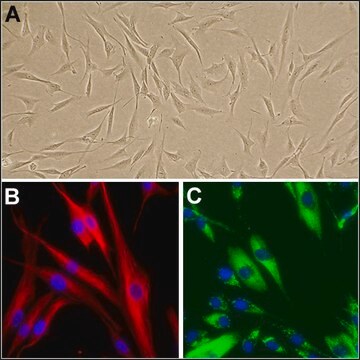
Cardiac fibroblasts are the most prevalent cell type in the heart, making up 60-70 % of all cells. HCF from Cell Applications, Inc. provide an excellent model system to study many aspects of human heart function and pathophysiology.
HCF has been utilized in a number of research publications, for example to:
- Determine that electrical coupling between cardiomyocytes and fibroblasts is mediated by large-conductance Ca2+-activated K+ channels that can be stimulated by estrogen receptor agonists (Wang, 2006, 2007); and show that antimitogenic effects of estradiol on HCF growth are mediated by cytochromes 1A1/1B1-and catechol-O-methyltransferase-derived metabolites (Dubey, 2005)
- Show that in response to mechanical stretch, cardiac fibroblasts release TGF-β which causes trombomodulin downregulation, increasing the risk of thromboembolic events (Kapur, 2007) and also induces cardiac fibroblast differentiation into myofibroblasts via increased Smad2 and ERK1/2 phosphorylation, that could be stimulated by endothelin-1 and inhibited by Ac-SDKP (Peng, 2010)
- Demonstrate that activation of G protein-coupled receptor kinase-2 (GRK2) prevents normal regulation of collagen synthesis in cardiac fibroblasts mimicking heart failure phenotype (D’Souza, 2011); identify FGF2 signaling pathway as potential target for modulating apoptosis in cardiac pathology (Ma, 2011) and investigate the roles of scleraxis (Bagchi, 2012) and AMPKα1 (Noppe, 2014) in scar formation following myocardial infarction
- Show that the KATP channel opener KMUP-3 preserved cardiac function after myocardial infarction by enhancing the expression of NO synthase and restoring MMP-9/TIMP-1 balance (Liu, 2011)
HCF were also shown to express delayed rectifier IK, Ito, Ca2+-activated K+ current (BKCa), inward-rectifier (Kir-type), and swelling-induced Cl- current (ICl.vol) channels (Yue, 2013).
Cell Line Origin
Application
Components
Preparation Note
- 1st passage, >500,000 cells in Basal Medium containing 10% FBS & 10% DMSO
- Can be cultured at least 8 doublings
Subculture Routine
Disclaimer
Storage Class Code
11 - Combustible Solids
WGK
WGK 3
Flash Point(F)
Not applicable
Flash Point(C)
Not applicable
Certificates of Analysis (COA)
Search for Certificates of Analysis (COA) by entering the products Lot/Batch Number. Lot and Batch Numbers can be found on a product’s label following the words ‘Lot’ or ‘Batch’.
Already Own This Product?
Find documentation for the products that you have recently purchased in the Document Library.
Customers Also Viewed
Protocols
Isolated from adult human heart ventricles, human cardiac fibroblasts (HCF) are used to study myocardial infarction, cardiac fibrosis, and cardiac hypertrophy. They play a critical role in extracellular matrix maintenance, growth factor synthesis, and cytokine synthesis. This protocol outlines the recommended procedure for culturing and subculturing human cardiac fibroblasts.
Our team of scientists has experience in all areas of research including Life Science, Material Science, Chemical Synthesis, Chromatography, Analytical and many others.
Contact Technical Service
