04942078001
Roche
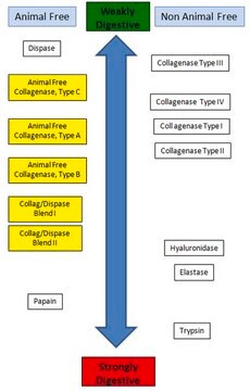
Dispase® II (neutral protease, grade II)
lyophilized, from bacterial, Roche, pkg of 5 × 1 g
Synonym(s):
dissociating enzyme, neutral protease, tissue dissociating
About This Item
Recommended Products
biological source
bacterial
Quality Level
sterility
non-sterile
form
lyophilized
specific activity
≥0.8 units/mg protein (37 °C, casein as substrate, pH 7.5)
packaging
pkg of 5 × 1 g
manufacturer/tradename
Roche
optimum pH
6.0-8.5
application(s)
sample preparation
Related Categories
General description
Specificity
Application
Features and Benefits
- Rapid, effective, yet gentle agent that liberates cells with minimal cell damage
- Maintains cell membrane integrity
- Non-mammalian source - free of mycoplasma and animal virus contamination
- Extremely stable to influences of temperature, pH, and interference by serum components
- Easily inactivated by chelating agents or by dilution
- Delivers higher activity and convenience
Unit Definition
A practical comparison of Roche units of Dispase® II with those cited in the Japanese literature (where Dispase® concentrations of 1,000 to 2,000 units/ml are not uncommon) suggests that one Roche unit equals approximately 600 Japanese units of dispase.
One unit of Roche Applied Science Dispase® equals 181 protease units (PU) measured as release of amino acids equivalent to 1 μg tyrosine per min and ml at pH 7.5 and +37 °C.
Preparation Note
Inhibitors: EDTA, EGTA, Hg2+, other heavy metals. Dispase is not inhibited by serum.
Working concentration: 0.6 to 2.4 U/ml
Working solution: Preparation of stock and working solutions:
To produce a 10 mg/ml stock solution, dissolve the lyophilized Dispase® II enzyme in HEPES-buffered saline (50 mM HEPES/KOH pH 7.4, 150 mM NaCl). To produce the working solution, dilute the above stock solution with the culture medium for the isolated cells, at a final concentration of 0.6 to 2.4 U/ml. Note that concentrations higher than 2.4 U/ml are not recommended. For best results, filter the working solution using a 0.22 μm filter membrane.
Storage conditions (working solution): -15 to -25 °C
The reconstituted stock solution is stable at 2 to 8 °C for 2 weeks. For storage up to 2 months the stock solution should be frozen in aliquots. Avoid repeated freezing and thawing! The working solution diluted with PBS is stable at 2 to 8 °C for 3 days.
Storage and Stability
Other Notes
Legal Information
Signal Word
Danger
Hazard Statements
Precautionary Statements
Hazard Classifications
Eye Irrit. 2 - Resp. Sens. 1 - Skin Irrit. 2 - STOT SE 3
Target Organs
Respiratory system
Storage Class Code
11 - Combustible Solids
WGK
WGK 1
Flash Point(F)
does not flash
Flash Point(C)
does not flash
Certificates of Analysis (COA)
Search for Certificates of Analysis (COA) by entering the products Lot/Batch Number. Lot and Batch Numbers can be found on a product’s label following the words ‘Lot’ or ‘Batch’.
Already Own This Product?
Find documentation for the products that you have recently purchased in the Document Library.
Customers Also Viewed
Articles
Enzyme Explorer Key Resource: Collagenase Guide.Collagenases, enzymes that break down the native collagen that holds animal tissues together, are made by a variety of microorganisms and by many different animal cells.
Related Content
Collagenase Guide.Collagenases, enzymes that break down the native collagen that holds animal tissues together, are made by a variety of microorganisms and by many different animal cells.
Our team of scientists has experience in all areas of research including Life Science, Material Science, Chemical Synthesis, Chromatography, Analytical and many others.
Contact Technical Service